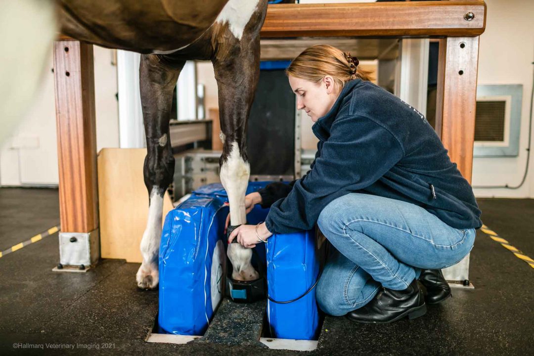


The use of veterinary MRI is growing exponentially, and it is considered the gold standard imaging modality for the evaluation of brain and spinal cord for small animals. Although radiographs and CT studies are able to help us evaluate the skeleton with phenomenal detail enabling detection of fractures, luxations, bone lysis and proliferation as well as acute disc disease, the evaluation of nervous system parenchyma can remain difficult or impossible, even when these studies are augmented by contrast agents.
Small Animal Brain Disease
Brain diseases, which can be detected utilising MRI and which require such a high level of imaging detail, include strokes, encephalitis, tumours, secondary consequences of seizure activity and congenital malformations. Looking a little further into a few of these specific brain diseases, the necessity of MRI can be expanded upon:
- Acute onset of brain disease may be due to one of several causes which include strokes. MRI is the only imaging modality which can definitely confirm the presence of this vascular crisis using the specialised sequences of diffusion weighting. An expedited accurate diagnosis of this disease can lead to a more comprehensive investigation of the underlying causes of stroke which can prevent further events in the future.
- Inflammations of the brain or encephalitis can be fatal, yet they may only cause minimal visible damage in the early stages. It is the early recognition of these inflammations that is vital for the patients to receive the accurate and aggressive treatment which they require to prevent a potential rapid deterioration and death. Only MRI has the capability to detect such early brain changes.
- Dogs and cats with seizures can have several possible brain diseases including tumours, stroke, inflammation and idiopathic epilepsy. The diagnosis of idiopathic epilepsy often requires the elimination of all other causes but itself may be associated with extensive oedema in the brain in the first few days and weeks after severe seizure activity; this can explain ongoing neurological dysfunction such as visual changes and behavioural abnormalities which otherwise might suggest a disease requiring a different treatment – MRI is essential to visualise these changes.
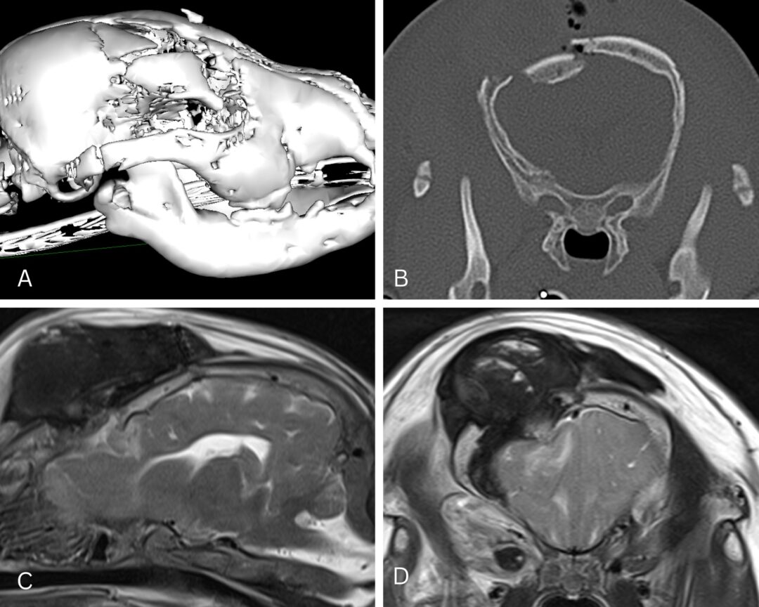
- The assessment of the ventricular system requires a 3D approach in many breeds which may have incidental enlargement of the ventricles, to determine what may be responsible for the clinical signs. Several characteristics of hydrocephalus have now been documented which absolutely require MR investigation and include periventricular hyperintensity, deformation of the overlying white matter and gyri and expansion of the lateral ventricles into the olfactory recesses. CT evaluation lacks the detail necessary to make this diagnosis and if it cannot be made, a plethora of other causes may have to be considered, often delaying the appropriate treatment required.
Diseases Affecting the Spinal Cord in Small Animals
Diseases affecting the spinal cord are not much different when it comes to the benefit of MRI. Most commonly we deal with disc disease, discospondylitis, neoplasia and congenital lesions such as syringomyelia affecting many of the Toy breeds, especially Cavaliers.
- Disc disease may be seen on CT if the extruded material is very mineralised but otherwise the essential 3D localisation of the spinal cord compression necessary for surgical planning requires MRI.
- Discospondylitis can be detected 1-2 weeks earlier than when using radiographs alone and if it extends into the spinal canal as empyema, it’s the only modality to provide the answer.
- Neoplasia affecting the spinal cord can come in many different forms and its precise location can be very helpful in determining what type of tumour it is, which translates into different treatment options and prognosis. No other imaging modality provides the essential and accurate soft tissue detail required for this.
- Lesions within the cord itself, such as the reasonably common toy breed disease syringomyelia, most often related to Chiari-like malformation, require a 3D soft tissue imaging investigation, an again MRI is essential for this purpose.
When looking for a rapid and effective imaging modality for many diseases affecting the nervous system, MRI is on top of the list.