

Coccidioidomycosis can sometimes be mistaken for a brain tumor in dogs, causing similar neurological signs and displays nearly identical, misleading characteristics on MRIs. The fungus often forms granulomas (nodules of inflammation) in the brain that mimic the appearance of glial tumors (gliomas) or other brain cancers.
The Patient
A 7-year-old female spayed Boxer dog mix presented for an acute onset of seizure activity, with 2 generalized tonic-clonic seizures noted by the owner in one day. During her initial hospitalization, she developed intermittent focal seizures described as fly-biting, starting the next day which progressed to focal motor status epilepticus. Once seizure activity was controlled, the dog was noted to pace in circles. Otherwise, the neurological examination was considered normal.
Thoracic radiographs revealed a tracheobronchial lymphadenopathy and a focal alveolar pattern in the right cranial lung lobe.
The dog underwent a brain MRI using the Hallmarq zero-helium, small animal, 1.5T machine (see Figs 1 & 2).
The Imaging Results
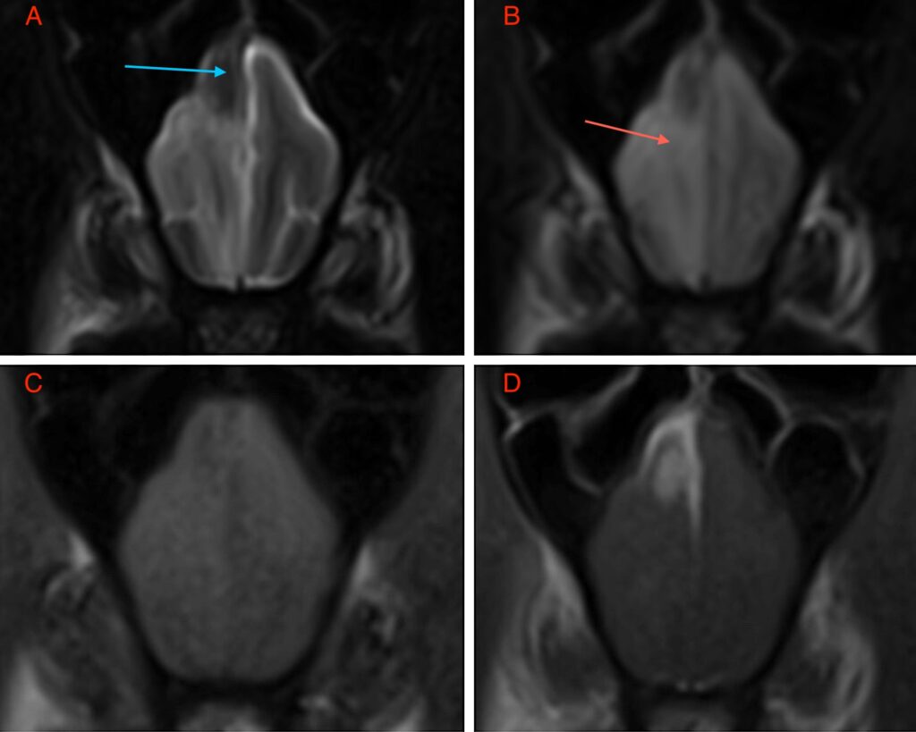
There is a fairly well-demarcated, T2W and FLAIR hypointense, T1W iso to hypointense and homogeneously contrast-enhancing extra-parenchymal lesion centered on the dorsal aspect of the right frontal lobe. The lesion does not exhibit T2* signal voids but there is a contrast-enhancing dural tail extending both ventrally down the midline falx and laterally along the dorsal surface. The mass measures 10mm x 6mm x 11mm. There is mild perilesional edema evident on T2W and FLAIR sequences which tracks along the white matter and causes mild mass effect. There is no evidence of transtentorial or cerebellar herniation.
The main differentials for this extra-parenchymal mass lesion include meningioma, histiocytic sarcoma, granular cell tumor and lymphoma, with meningioma being the most common possibility, although inflammatory diseases, of infectious and immune-mediated aetiology, can never be ruled out.
A CSF tap was unremarkable which can be compatible with a neoplastic disease rather than an inflammatory disease. However, infectious disease serology results confirmed positive IgG and IgM antibodies for Coccidioides. A pyogranulomatous encephalitis was then reported based on histopathology of lesion following a craniotomy and biopsy. The dog was treated for Coccidioides with fluconazole and is stable 4 months post-presentation.
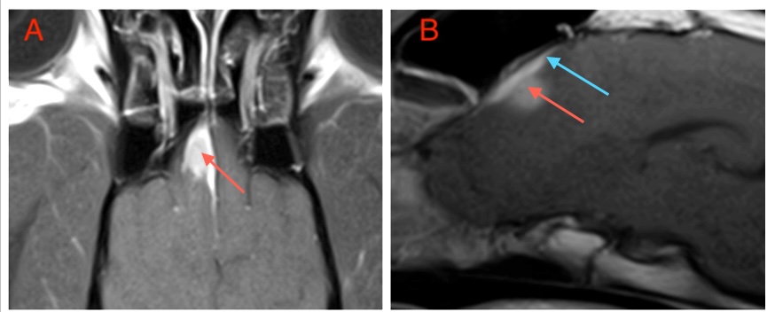
The Most Valuable MRI Sequence for This Case
The T1W post-contrast sequence demonstrates both meningeal involvement and the homogenous enhancement often seen associated with tumours such as meningioma originating outside of the blood-brain barrier but also with some granulomas. Typically, the extent of disease can be accurately assessed as the tissue which enhances. The extension of the mass lesion along the meninges as a ‘dural tail’ can be seen clearly in Fig 2B and is often a characteristic of meningiomas, although not 100% specific for this tumour type as seen with this case.
If your looking to provide an advanced imaging service to your pet owners, why not get in touch? Zero-helium small animal 1.5T MRI could be the solution…
Coccidioidomycosis: A Disease Overview
Coccidioidomycosis infections are most common in arid and semiarid regions of the southwestern US, as well as in similar areas of Mexico, Central, and South America, though cases have been reported in nonendemic regions. Many animal species, including humans, are susceptible, with dogs most frequently showing clinical signs. The infections are caused by Coccidioides immitis and C. posadasii. Infection typically occurs through inhalation of fungal arthrospores, which can be carried on dust particles. Epidemics may happen when rainy periods follow droughts, leading to dust storms.
The disease spectrum ranges from no symptoms to progressive, disseminated, and fatal cases. Primarily a respiratory disease, it can be self-limiting or become chronic. Dissemination occurs in about 20% of canine cases, affecting tissues such as the brain, eyes, skin, bones, and joints. Many dogs with disseminated disease show signs across multiple organ systems.
Clinical symptoms vary greatly depending on the infected organ and infection severity. Pulmonary-only cases often present with cough, lethargy, inappetence, fever, and rapid breathing. Dogs with central nervous system involvement may have seizures; other signs include vestibular symptoms, asymmetric cranial nerve deficits, behavioral changes, and circling.
A tentative diagnosis can be made based on positive serology combined with consistent clinical signs, but some animals without symptoms may also test positive due to prior exposure or subclinical infection.
Serologic testing for antigens has been largely insensitive, although recent studies show EIA antigen testing of CSF can be positive in some cases. EIA antibody testing of CSF may also be beneficial, but some patients with brain disease unrelated to coccidioidomycosis may also test positive.
MRI and it’s use in Diagnosis
MRI can assist in diagnosing CNS coccidioidomycosis. Most cases display a single, contrast-enhancing granuloma with surrounding edema in the cerebrum. Differentiating these from other masses is challenging. A second pattern involves bilateral encephalitis of the frontal lobes and caudate nuclei, evident on T2 images. These cases may resolve clinically but often show marked atrophy of affected brain regions.
While the disease can resolve on its own, long-term antifungal therapy is necessary if chronic respiratory, neurological, or multisystemic signs are present. Avoid compounded azole medications, as they may lack adequate therapeutic doses.
Fluconazole (5–10 mg/kg daily) is the most common treatment. Itraconazole (10 mg/kg daily) is also used, especially in cases involving bones, though it can be more expensive and cause more side effects. Investigations suggest that compounded itraconazole from bulk drug is ineffective and should not be used for serious infections.
With thanks to Dr. Casey Birkel DACVIM-Neuro at Tucson Veterinary Specialists for sharing this case study with us.
References
- Bentley RT, Heng HG, Thompson C, Lee CS, Kroll RA, Roy ME, Marini L, Heo J, Wigle WL. MAGNETIC RESONANCE IMAGING FEATURES AND OUTCOME FOR SOLITARY CENTRAL NERVOUS SYSTEM COCCIDIOIDES GRANULOMAS IN 11 DOGS AND CATS. Vet Radiol Ultrasound. 2015 Sep-Oct;56(5):520-30
- Davidson AP, Shubitz LF, Alcott CJ, Sykes JE. Selected Clinical Features of Coccidioidomycosis in Dogs. Med Mycol. 2019 Feb 1;57(Supplement_1):S67-S75
- Spoor E, Stainback L, Plummer S, Knowles K. A novel form of intracranial coccidioidomycosis is present in dogs and exhibits characteristic clinical and magnetic resonance imaging findings. Vet Radiol Ultrasound. 2019 Jan;60(1):47-55
