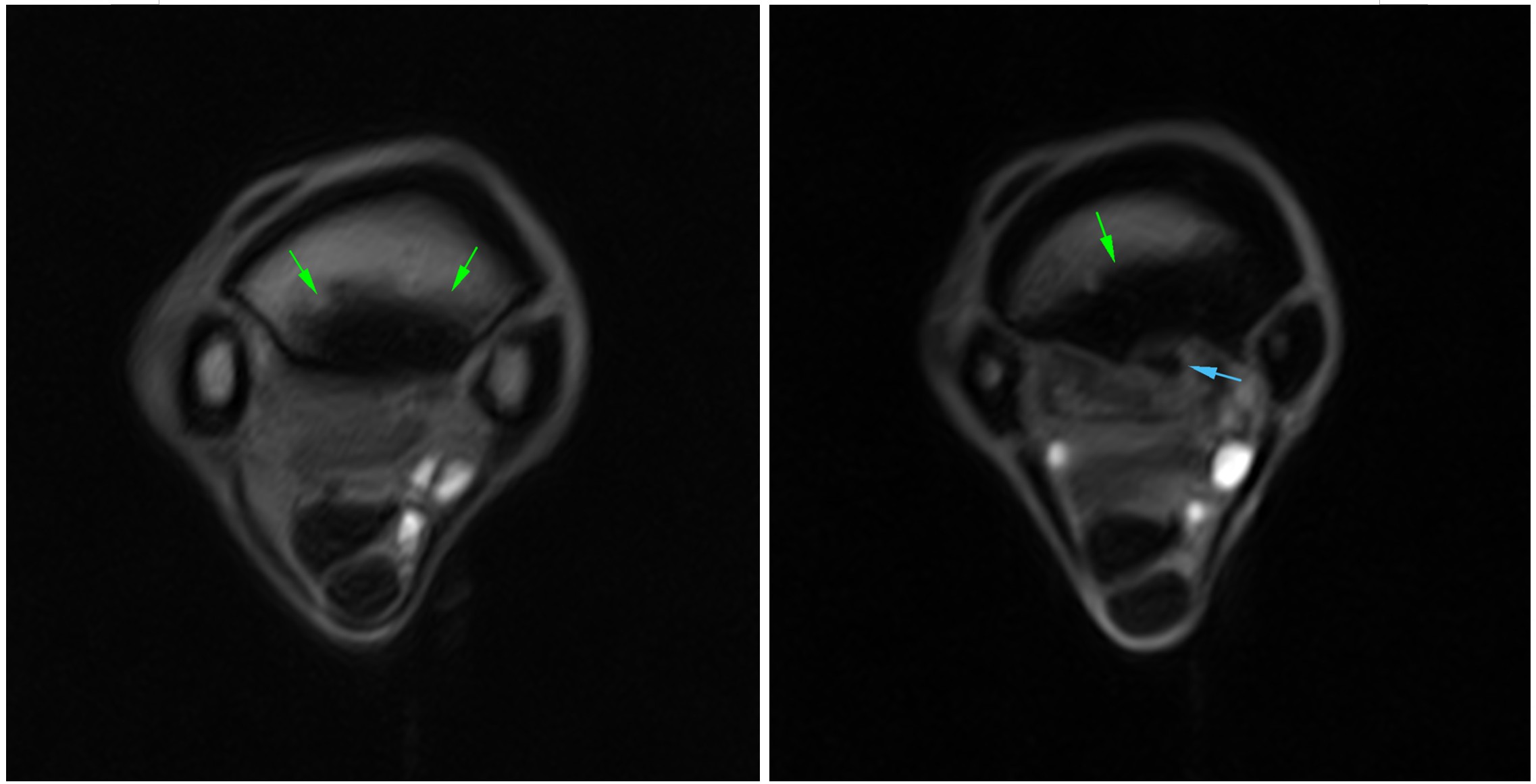
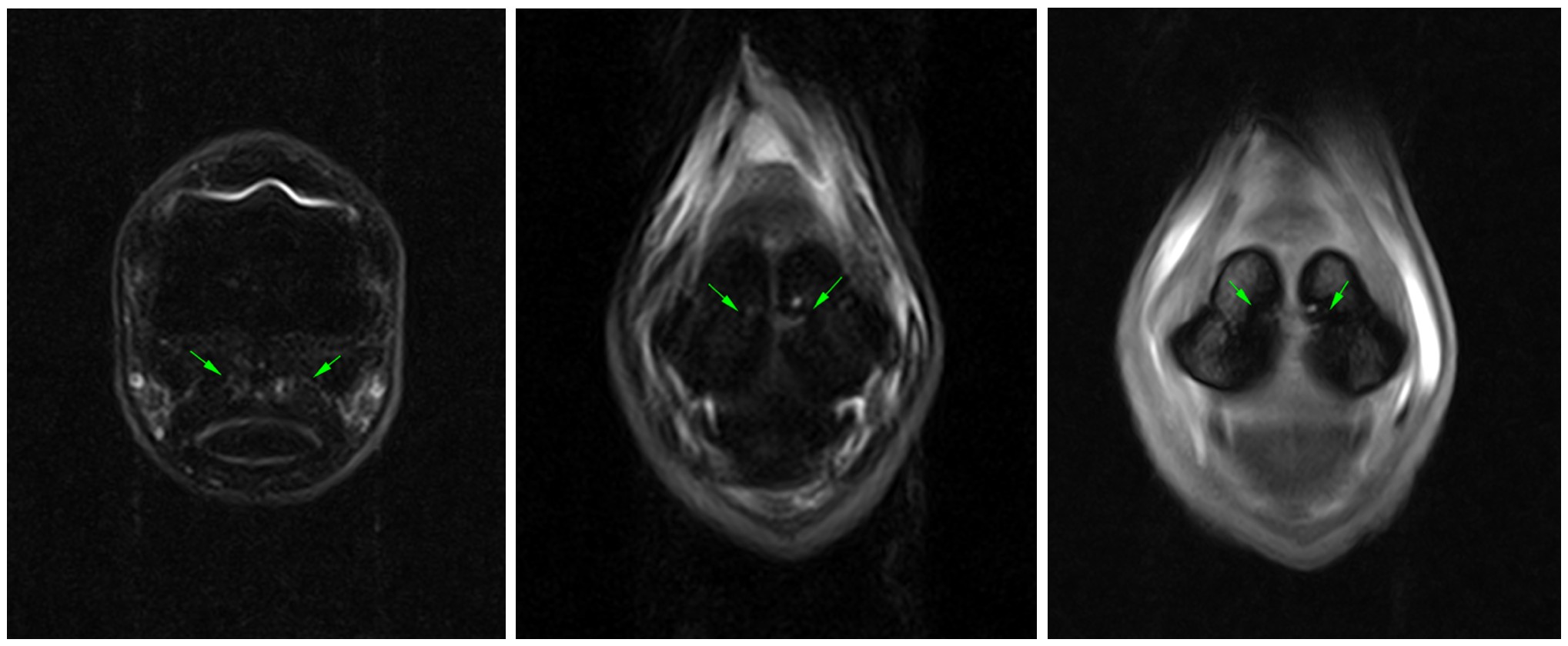
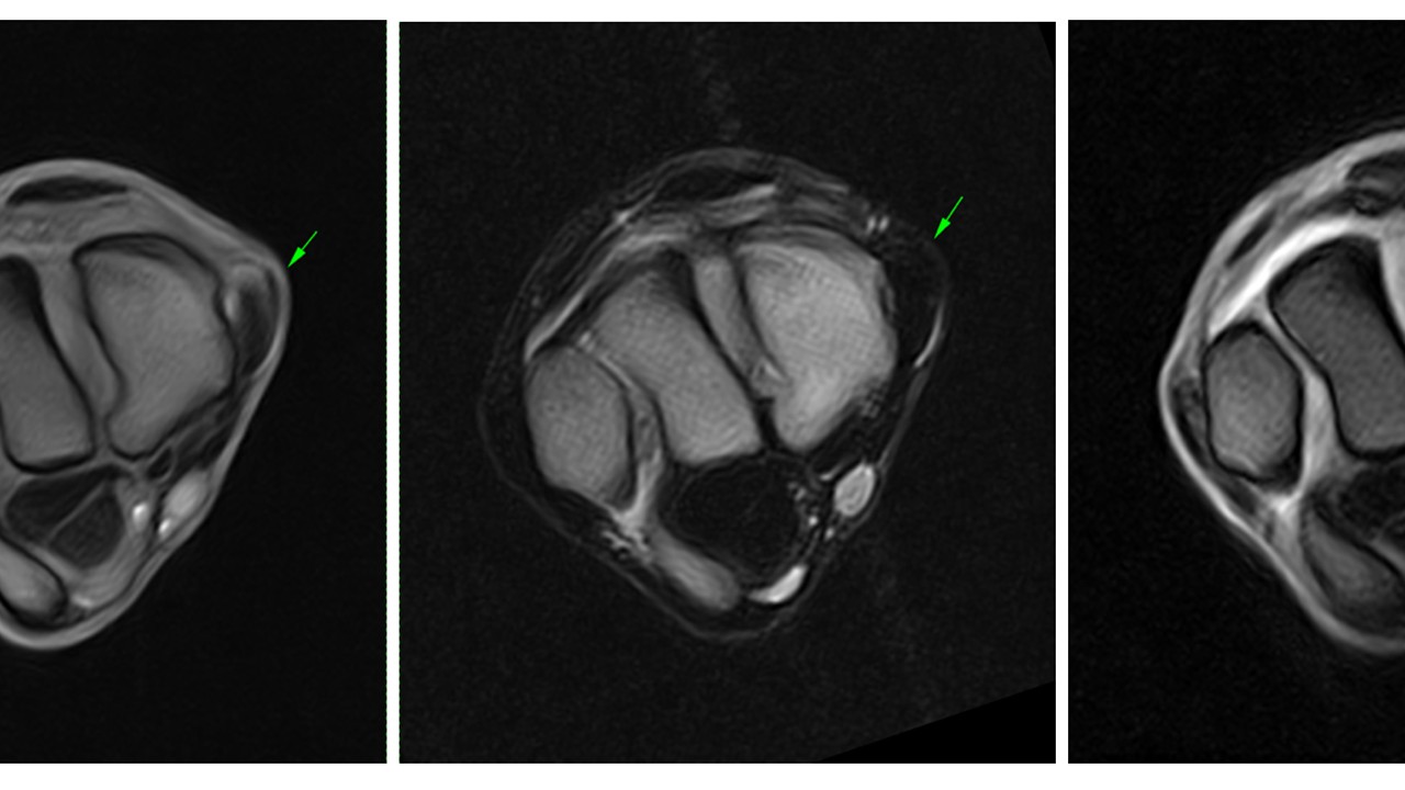
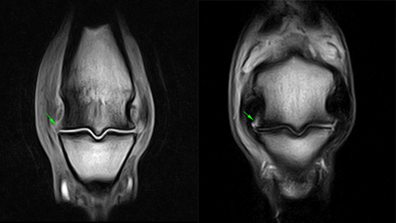
Skip to the content

Looks like you’re on our UK site! Would you like to switch US site?

Do not ask again
Switch to US

Welcome to the next generation!

Introducing the World’s First Veterinary-Specific Zero-Helium (ZHM) Small Animal 1.5T MRI

Find out more
Header image

Region

  • English (USA)
    • English (UK) (English)
Community
Hallmarq Veterinary Imaging
Innovative diagnostic imaging solutions
  • Products & Services
    • Standing Equine MRI
    • Standing leg CT Scanner for Horses
    • Small Animal MRI
  • Solutions
    • Q-Care
    • MRI – for Equine Vets
    • MRI – for Horse Owners
    • CT – for Equine Vets
    • CT – for Horse Owners
    • For Small Animal Vets
    • For Pet Owners
  • News
  • Resources
    • Case Studies
    • Webinars
    • Download Center
  • About
    • About us
    • Meet the team
    • Find a Hallmarq Site
    • Environmental, Social and Governance (ESG)
  • Contact us
Search
Menu

Case Studies

Explore current trends and benefit from our users’ clinical results
Close search
Close
  • Products & Services
    Show sub menu
    • Standing Equine MRI
    • Standing leg CT Scanner for Horses
    • Small Animal MRI
  • Solutions
    Show sub menu
    • Q-Care
    • MRI – for Equine Vets
    • MRI – for Horse Owners
    • CT – for Equine Vets
    • CT – for Horse Owners
    • For Small Animal Vets
    • For Pet Owners
  • News
  • Resources
    Show sub menu
    • Case Studies
    • Webinars
    • Download Center
  • About
    Show sub menu
    • About us
    • Meet the team
    • Find a Hallmarq Site
    • Environmental, Social and Governance (ESG)
  • Contact us

Region

  • English (USA)
    • English (UK) (English)
Community
  • All case studies
  • Equine MRI
  • Small Animal MRI
  • Equine CT

Hyperextension of the right front metacarpophalangeal joint

An 11-Year-Old Female Spayed Poodle Presented for an Onset of Seizure Activity

slCT Case Study TeleMed Vet

A Substantial POD Lesion With Mild Uptake on Scintigraphy Images

A left hind limb lameness resulting from a thrombosed digital artery

Thoroughbred mare with an acute exacerbation of an intermittent low-grade right hindlimb lameness

SAMRI Case Study #4

The use of MRI in Detecting Spina Bifida of the Canine Sacrum.

Hypertrophic Meningitis

Hypertrophic Meningitis

Intracranial Intra-arachnoid Diverticulum

Cerebral Microbleeds

Standing Equine Leg CT Clinical Images Gallery

A horse being scanned by the equine MRI machine

10-year-old Irish Sports horse with an inflammatory bone lesion in the pastern

Horse in Equine MRI scanner

Investigation of an osteolytic lesion on the medial border of the distal phalanx

Next page →
Home > Case Studies

Keep up to date

Improving the lives of animals and the people who care for them.

Email

email hidden; JavaScript is required

Follow Us

  • Facebook
  • LinkedIn
  • Twitter

Product & Services

  • Standing Equine MRI
  • Standing leg CT Scanner for Horses
  • Small Animal MRI

Solutions

  • Q-Care
  • MRI – for Equine Vets
  • MRI – for Horse Owners
  • CT – for Equine Vets
  • CT – for Horse Owners
  • For Small Animal Vets
  • For Pet Owners

About

  • About us
  • Meet the team
  • Find a Hallmarq Site
  • Environmental, Social and Governance (ESG)

© Hallmarq Veterinary Imaging 2023

Registered in 1275 W. Roosevelt Rd., Suite 116, West Chicago, IL 60185, USA

  • Privacy Policy
  • Cookie Policy
  • Terms of Use
  • Sitemap

Web design Surrey