

In this case from Liphook Equine Hospital, the identification of superficial digital flexor tendinopathy within the carpal sheath was an unusual finding but was considered the primary contributor to the lameness. We find out out imaging with standing MRI made the difference to diagnosis.
History
The horse was reported to have recurrent left forelimb lameness, which showed partial improvement following intra-articular anesthesia of the middle carpal joint and a lateral palmar nerve block.
MRI Findings
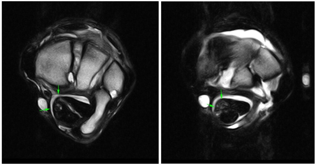
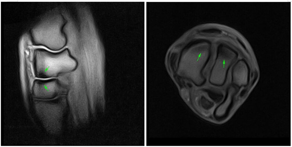
The horse underwent standing MRI of the carpus and proximal suspensory. This revealed irregular margin of the superficial digital flexor tendon (SDFT) within the carpal sheath with loss of definition of its lateral margin. There was moderate soft tissue proliferation within the medial aspect of the carpal sheath and loss of differentiation between the medial margin of the deep digital flexor tendon (DDFT) and the SDFT, extending to the level of the carpometacarpal joint. The carpal sheath was moderately effused.
There was also mild/moderate thickening of the distal subchondral bone plate of the radial carpal bone and also of the opposing surface of the radial facet of the carpal bone. Similar thickening was at the medial aspect of the intermediate carpal bone and intermediate facet of the third carpal bone. There was moderate effusion of the middle carpal joint and mild periarticular new bone formation at the periphery of the joint.
Conclusion
The MRI findings were consistent with mild superficial flexor tendinopathy within the carpal sheath, with moderate effusion and soft tissue proliferation within the sheath. Additional findings included mild adaptive modelling and osteoarthropathy of the middle carpal joint, with associated synovitis.
The identification of superficial digital flexor tendinopathy within the carpal sheath was an unusual finding and was considered the primary contributor to the lameness.
The use of iNAV motion correction – together with AI applications such as PAICE – was important in this case. When combined, both help to reduce scan times and improve image quality when scanning the proximal limb and therefore supporting an accurate diagnosis.
With thanks to Dr Lucy Meehan-Howard and Suzy Hall, Liphook Equine Hospital, UK for sharing this case with us.