


Abstract
In this study, we take an in-depth look into the use of Magnetic Resonance Imaging (MRI) as a diagnostic tool in a complex multi-limb lameness in an event horse. A 10-year-old Irish Sport Horse mare actively competing in affiliated eventing was presented for investigation of a complex and evolving lameness syndrome following the acute onset of severe right forelimb lameness during competition.
Initial clinical assessment, diagnostic analgesia, radiography, and ultrasonography identified bilateral forefoot pain, proximal suspensory desmopathy, and mild osteoarthritic change within the distal tarsal joints. However, the severity and fluctuating nature of the clinical signs remained difficult to reconcile with the diagnostic findings.
Subsequent low-field standing magnetic resonance imaging (MRI) identified significant soft tissue pathology within the right forefoot, including:
- deep digital flexor tendon injury
- collateral sesamoidean ligament pathology
- navicular bone abnormalities
- bilateral distal interphalangeal joint synovitis
MRI findings provided a coherent explanation for the unusual clinical presentation, enabled refinement of the treatment plan, and facilitated improved owner understanding and engagement. This case highlights the diagnostic value of standing MRI in horses with complex multi-limb lameness patterns where conventional imaging modalities fail to adequately explain clinical findings.
Introduction
Lameness in performance horses frequently presents a diagnostic challenge, particularly when multiple concurrent pathologies are present. Compensatory gait adaptations may mask the primary source of pain and exacerbate subclinical disease processes, resulting in complex presentations that are difficult to interpret using conventional diagnostic techniques.
Magnetic resonance imaging has become an increasingly important tool for investigating foot-related lameness due to its ability to characterize both osseous and soft tissue structures. This report describes the diagnostic investigation and clinical management of a competition horse presenting with acute onset forelimb lameness superimposed upon previously unrecognized chronic musculoskeletal disease.
Case Presentation
A 10-year-old Irish Sport Horse mare used for affiliated eventing was referred for lameness investigation. The horse had no previous history of clinically significant lameness and had been competing successfully prior to presentation.
One month before referral, the mare developed acute severe right forelimb lameness, graded 9/10, while cantering in a competition warm-up arena. Clinical signs improved rapidly, and lameness became mild at the walk. The horse was considered fit for transportation by attending veterinarians and returned home, where paddock rest and oral phenylbutazone therapy were instituted for one week.
Subsequent assessment by a RAMP-registered physiotherapist identified mild right forelimb and bilateral hindlimb lameness, accompanied by moderate generalized muscular spasm affecting the right cervical base, shoulder, and pectoral musculature. Veterinary assessment was therefore recommended.
Clinical Examination
Static examination revealed marked pain on palpation of the thoracic region immediately caudal to the withers, particularly on the right side. Saddle assessment demonstrated poor fit, with evidence of significant pressure and pinching over the withers.
Dynamic examination demonstrated:
- • 3/10 left hindlimb lameness in a straight line
- • 2/10 left forelimb and 3/10 left hindlimb lameness on the left rein
- • 1/10 right forelimb and 5/10 left hindlimb lameness on the right rein
Distal and proximal forelimb flexion tests were negative. Mild positive responses were observed following both distal and proximal hindlimb flexion tests.
Diagnostic Analgesia
A left forelimb palmar digital nerve block resulted in persistence of right forelimb lameness, graded 1/10 in a straight line, 2/10 on the left rein, and 4/10 on the right rein.
Following resolution of local anesthesia, blockade of the left distal interphalangeal (DIP) joint resulted in a transition to a milder right forelimb lameness pattern.
In the hindlimbs, a left low six-point nerve block reduced lameness to 2/10 in a straight line and on the left rein, and 3/10 on the right rein. Sequential blockade of the deep branch of the lateral plantar nerve further reduced lameness to 1/10 in the right hindlimb in a straight line and on the left rein, and 1/10 in the left hindlimb on the right rein. Intra-articular anesthesia of the stifle joint produced no appreciable improvement.
Diagnostic Imaging
Radiography
Radiographs of both forefeet demonstrated subtle modelling of the extensor process of the distal phalanx (P3) and the dorsodistal aspect of the middle phalanx (P2). Multiple radiolucent channels were present along the distal border of the navicular bone. These changes were bilaterally symmetrical and considered of equivocal clinical significance for a horse of this age, breed, and athletic discipline.
Radiographs of the tarsal region revealed narrowing of the distal intertarsal and tarsometatarsal joints, accompanied by mild osteophyte formation at the dorsolateral aspect of the tarsometatarsal joint. These findings were similarly considered of uncertain clinical importance. Heterogeneous radiolucency was also identified within the proximal third metatarsal bone.
Lateromedial radiographs of the hind feet demonstrated a marked negative plantar angle of P3. Additional modelling of the extensor process of P3 and dorsodistal P2, particularly within the left hind foot, was observed.
Ultrasonography
Ultrasonographic examination of the proximal suspensory ligaments identified enlargement of ligament cross-sectional area bilaterally, greater in the right hindlimb than the left. Heterogeneous echogenicity, including regions of fibre disruption and fibrosis, was evident on transverse views.
Longitudinal imaging demonstrated modelling of the proximal third metatarsal bone and loss of normal fibre architecture at the bone-ligament interface, particularly within the left hindlimb.
Initial Diagnosis and Management
Based on the available diagnostic findings, primary diagnoses of bilateral forefoot pain, proximal suspensory desmopathy, and osteoarthritis of the distal tarsal joints were established.
The owner found these diagnoses difficult to reconcile with the horse’s previous athletic performance and the sudden onset of severe right forelimb lameness. It was hypothesized that acute soft tissue injury within the right forelimb had induced compensatory biomechanical adaptations, resulting in exacerbation of pre-existing but clinically silent musculoskeletal abnormalities.
Owing to financial considerations and the evolving clinical picture, conservative management was elected. This consisted of remedial hindlimb farriery using quarter-clipped shoes with neutral 3D half-mesh pads, restricted turnout, and a month of controlled straight-line walking exercise. Further investigation of the forefeet was deferred pending reassessment.
Clinical Deterioration
Upon re-examination after one month, the horse exhibited worsening left hindlimb lameness shortly after remedial farriery was implemented. Elevated digital pulses were present in the left hind foot in the absence of focal hoof tester sensitivity.
Lameness was graded as:
- 5/10 in the left hindlimb in a straight line
- 7/10 on the left rein
- 5/10 on the right rein
Palmar digital nerve blockade resulted in considerable improvement, reproducing the milder lameness pattern observed initially. Follow-up radiographs excluded obvious fracture pathology. The episode was considered most consistent with nail bind, and treatment consisted of shoe removal, foot poulticing, and oral non-steroidal anti-inflammatory medication.
Two weeks later the mare was reassessed following replacement of the shoe. Digital pulses had normalized; however, dynamic examination identified continued right forelimb and left hindlimb lameness. Repeat diagnostic analgesia localized pain once again to the right forefoot and to both the left hind foot and proximal suspensory region.
At this stage, the fluctuating nature of the forefoot pain was increasingly difficult to explain solely on the basis of previous diagnostic findings, prompting advanced imaging.
MRI Findings
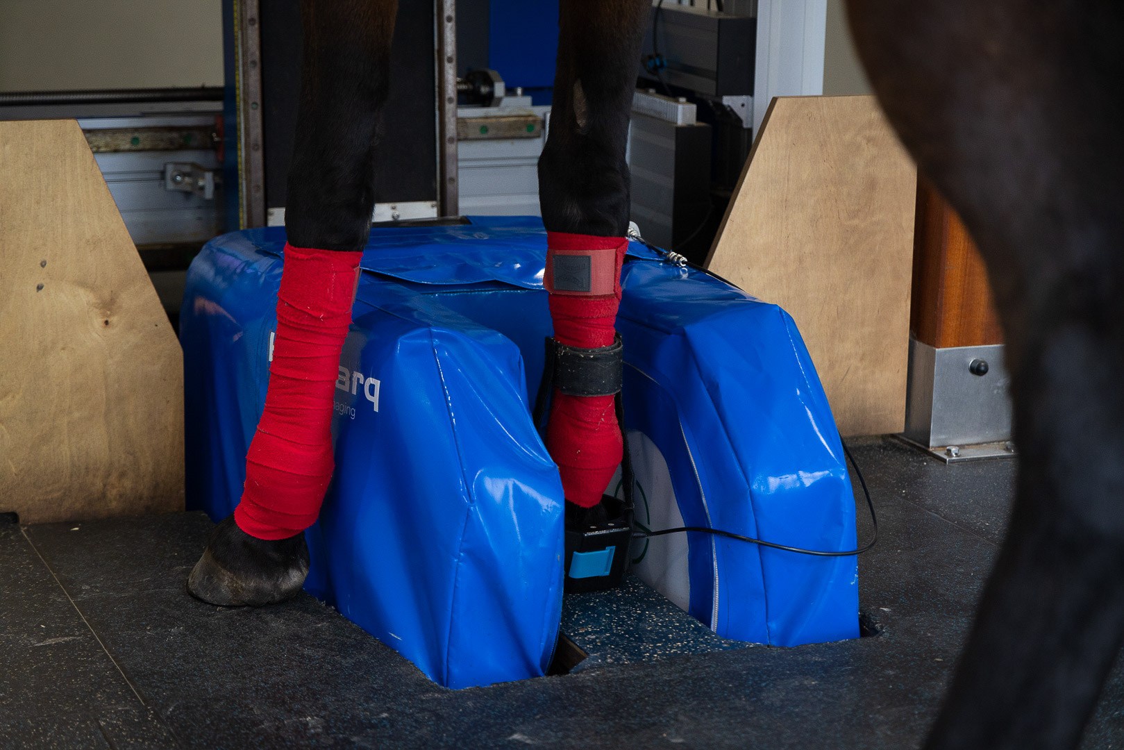
Standing low-field MRI was performed using a Hallmarq 0.27T MRI machine. Image interpretation by M. Smith, Image Equine Ltd, identified the following abnormalities:
Right Forefoot
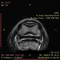
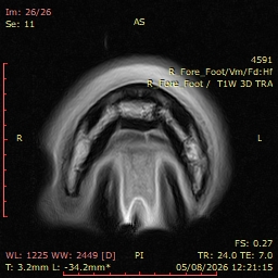
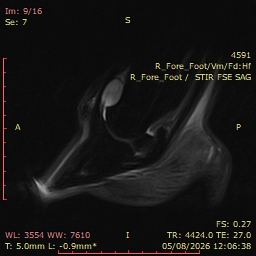
See Figs. 1, 2 and 3 below.
- Linear tendonitis affecting the medial lobe of the distal deep digital flexor tendon (DDFT)
- Mild DDFT tendonitis within the proximal recess of the navicular bursa
- Adhesion formation between the DDFT and collateral sesamoidean ligament (CSL)
- Collateral sesamoidean ligament desmitis
- Diffuse inflammatory and degenerative changes within the navicular spongiosa
- Moderate distal interphalangeal joint synovitis and periarticular remodelling
Left Forefoot
See Fig. 4 below.
- Moderate distal inte Mild DDFT tendonitis within the proximal recess of the navicular bursa
- Adhesion formation between the DDFT and collateral sesamoidean ligament (CSL)
- Collateral sesamoidean ligament desmitis
- Moderate distal interphalangeal joint synovitis and periarticular remodelling
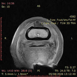
Enlargement of the dorsal margin of the DDFT within the proximal recess of the navicular bursa.
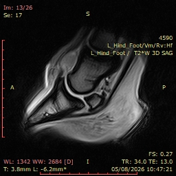
Left Hindfoot
See Fig. 5 below.
- Significant distal interphalangeal joint synovitis
- Mildly broken-back hoof-pastern axis
Discussion
MRI findings strongly suggested that the initial acute lameness episode was attributable to significant injury of the medial lobe of the right forelimb DDFT. The severity of this injury was not apparent using conventional diagnostic techniques. Increased weight-bearing by the contralateral forelimb likely exacerbated pre-existing chronic bilateral foot pathology, resulting in the fluctuating bilateral forelimb lameness observed throughout the investigation.
Importantly, MRI excluded significant primary pathology within the left hind foot and provided evidence supporting a mechanically induced inflammatory flare-up of the distal interphalangeal joint following remedial farriery. Although unilateral deterioration following bilateral farriery intervention is unusual, the pre-existing radiographic abnormalities and more severe proximal suspensory pathology affecting the left hindlimb provide a logical explanation.
The MRI findings therefore allowed the disparate clinical signs, diagnostic analgesia results, and imaging findings to be integrated into a coherent diagnostic framework. This significantly improved owner understanding and facilitated acceptance of the revised treatment strategy.
Treatment and Prognosis
Following MRI diagnosis, remedial hindlimb farriery was discontinued and the horse returned to barefoot management of the hind feet. Although this approach may increase mechanical strain upon the suspensory apparatus in horses with a marked negative plantar angle, alleviation of patient discomfort was prioritised. Intra-articular corticosteroid treatment of the left hind DIP joint remains a potential option should clinical improvement prove inadequate.
Given the confirmed right forefoot soft tissue injury and lack of consistent improvement during turnout-based rest, the horse was placed on strict box rest combined with a progressively increasing six-week walking exercise programme. Egg-bar shoes were applied to both forefeet to provide additional support.
Future therapeutic options include corticosteroid medication of the distal interphalangeal joints to address persistent synovitis and inflammation of the palmar foot structures. Subsequent therapies, including orthobiologic treatments or polyacrylamide hydrogel injections, may be considered to support long-term joint health.
Given the presence of multiple concurrent pathologies affecting several limbs, a guarded prognosis for return to and maintenance of soundness at affiliated eventing level has been provided. Should forelimb lameness resolve satisfactorily, surgical treatment of chronic proximal suspensory disease may be considered. However, the welfare implications and ethical considerations associated with returning such a horse to a jumping discipline would require careful evaluation.
Conclusion
This case demonstrates the considerable diagnostic value of standing MRI in horses presenting with complex, multi-limb lameness. Conventional diagnostic imaging and nerve blocks identified several clinically relevant abnormalities but failed to fully explain the severity and fluctuation of clinical signs. MRI successfully identified substantial right forefoot soft tissue pathology while excluding significant disease within the left hind foot, thereby enabling a comprehensive understanding of the lameness pattern and guiding appropriate treatment decisions.
With our thanks to Nikki Platt BVSc MRCVSand team at Oakhill Equine Vets, UK for sharing this study.