

Foreign Body at the White Line
In this case study, kindly provided by Valley Equine Hospital, UK, we examine a horse presenting with acute onset of right forelimb lameness. The horse exhibited a horizontal crack in the proximal lateral hoof wall near the white line, that intermittently discharged purulent material.
Radiographic Findings
Radiographs performed on two different occasions suggested the presence of either a foreign body or a small bony fragment adjacent to the distal phalanx.


Figure 1: Dorsopalmar and Figure 2: Dorsoproximal-palmarodistal oblique of the distal phalanx highlighting the suspected bony fragment. Lateral is to the left.
CT Findings
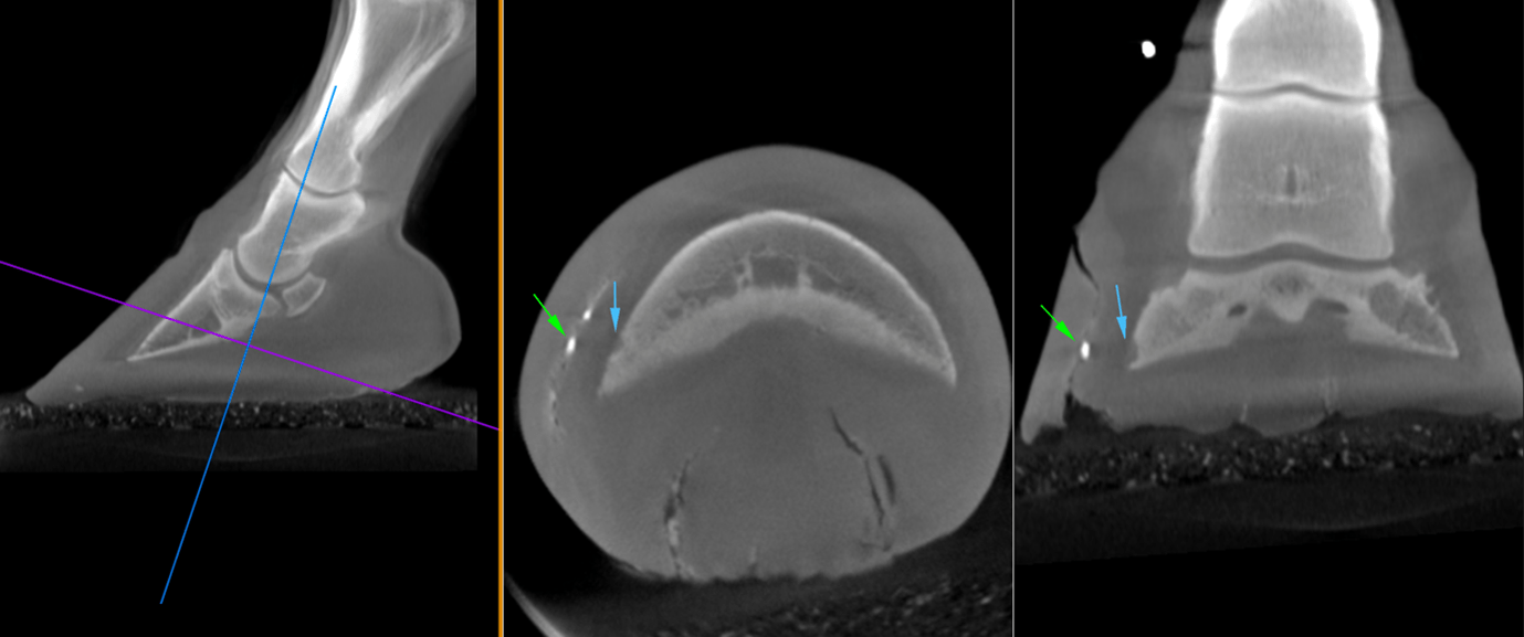
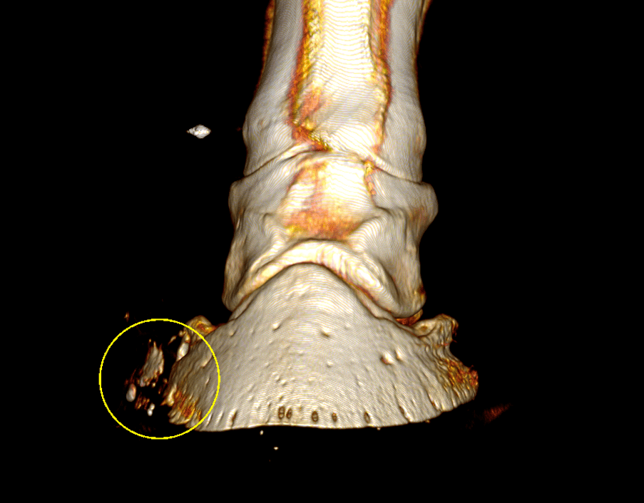
Imaging with Hallmarq’s Vision CT revealed that what appeared to be a small bony fragment on radiographs, was more consistent with metallic artefact. This artefact was noted in the white line approximately 6cm proximal to the level of the sole. The entire white line on the lateral aspect of the sole was separated. This extended proximally to communicate with the horizontal defect noted grossly on the proximal lateral aspect of the dorsal hoof wall. Adjacent to the metallic artefact was a small concave defect in the distal phalanx however, there did not appear to be further change to the distal phalanx consistent with septic pedal osteitis.

Surgery
Based on the CT findings the horse underwent standing surgery. The entire lateral aspect of the white line was opened and debrided. The region that revealed the metallic object and adjacent small change in the distal phalanx on CT scan, was the site for the most widespread and aggressive debridement.
With many thanks to Dr Tom McParland BVetMed DiplACVS (LA) MRCVS and Melissa Lockwood RVN, Valley Equine Hospital, UK, for sharing this case with us.