

History
In this multimodal imaging case, a 15-year-old Quarter Horse gelding, used for barrel racing, was presented with mild left hind limb lameness. The tarsal joints were initially treated, however, two weeks later, following a barrel race, the lameness recurred. Scintigraphic and radiographic examinations were performed, followed by Magnetic Resonance Imaging (MRI). A central tarsal bone fracture was then diagnosed which enabled a treatment plan using CT-guided screw fixation.
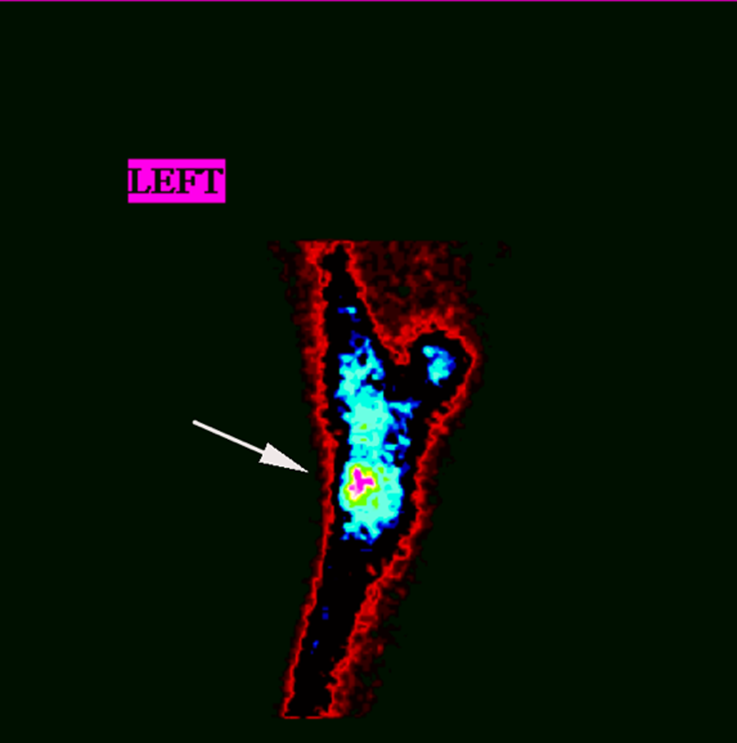
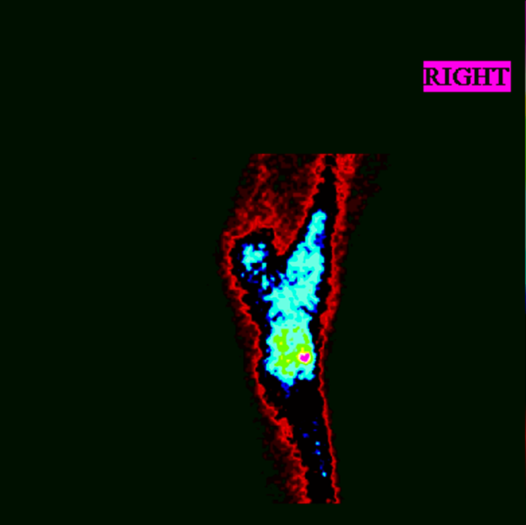
Scintigraphy Findings
Initially, the horse underwent scintigraphic examination, which revealed moderate focal increased radiopharmaceutical uptake (IRU) in the central tarsal bone on the lateral views of the left tarsus (Figs.1A and 1B).
Radiographic Findings
Radiographic examination showed no detectable abnormalities. The distal intratarsal joint was medicated, and the horse resumed racing. However, within few days, it developed a 3/5 left hindlimb lame.
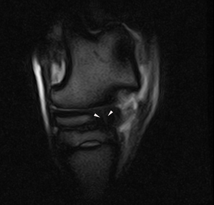
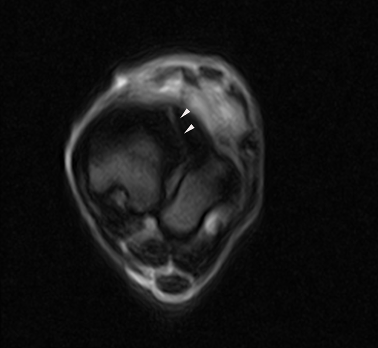
MRI Findings
Standing Equine MRI of the left tarsus was elected and revealed a bi-articular fracture of the central tarsal bone that extended in a dorso-medial to plantar-lateral direction through the lateral third of the bone. The fracture had sharp margins, was surrounded by moderate sclerosis, and was minimally displaced in an abaxial direction (Figs. 2A and 2B).
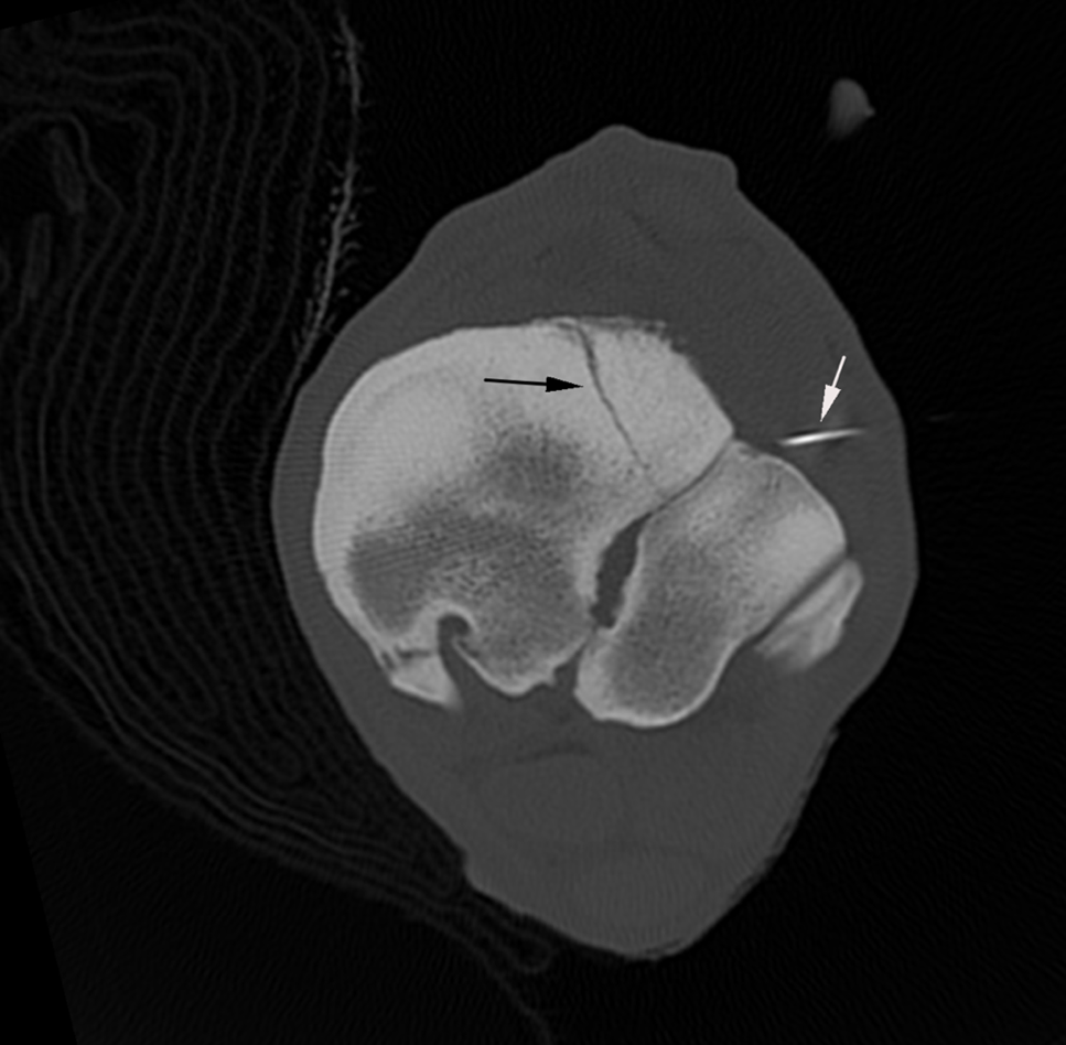
CT Guided Screw Fixation
The horse underwent general anesthesia, and internal fixation was achieved with the placement of a screw (Fig. 3) under CT guidance.
Conclusion
Multimodal imaging in this case, led to a definitive diagnosis and appropriate treatment. The horse recovered well and successfully returned to competitive barrel racing 9 months after the initial injury.
With thanks to Dr C. Renee Andrea, Chaparral Veterinary Medical Center, Cave Creek US, for sharing this case study with us.
