Looks like you’re on our UK site! Would you like to switch US site?

Do not ask again

FAQ’s

Why refer for standing MRI?

Much has been learned about the causes of equine lameness since the advent of MRI.  From the previously under-diagnosed, such as collateral desmitis of the distal interphalangeal joint, through the previously misunderstood, such as navicular syndrome, to the previously unknown, such as bone marrow oedema, MRI has revolutionised our ability to provide a diagnosis and improve prognosis in equine lameness.

MRI is unparalleled in providing images of both soft and bony tissues.  Distinguishing water from fat, it highlights areas of pathology such as inflammation and bruising, in a way that radiography, CT, ultrasound or nuclear scintigraphy just can’t do.  By imaging the region of interest in slices orientated in any 3D plane, a lesion can be visualised without superimposition of adjacent structures.  Multiple views allow you to appreciate the full extent of the injury.
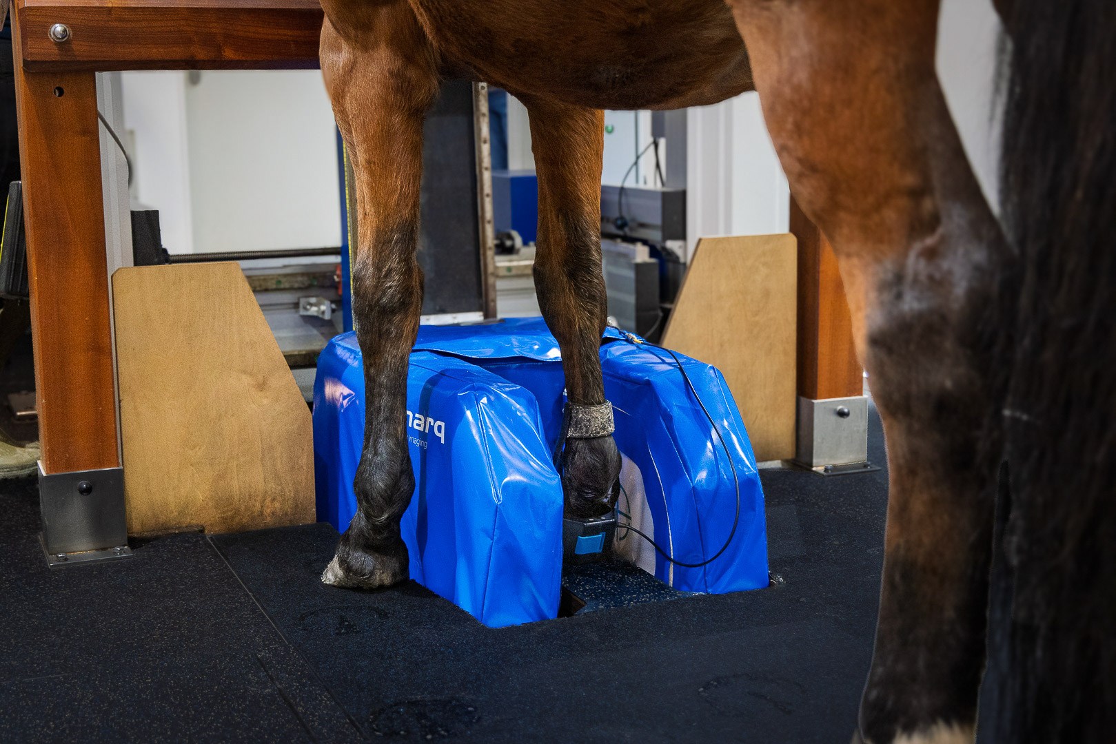
When would I refer for standing MRI?

It is important to localise the source of lameness before referring a case.  In particular, the abaxial sesamoid block alone is not sufficient. Horses referred for a foot scan should have a positive response to a palmar digital or coffin joint block.  Additional scans of the pastern and fetlock should be considered for horses that are positive to abaxial sesamoid but negative to more distal blocks.  You should ideally consider an MRI referral in the following cases:

  • Lameness localised by nerve blocks to a specific region the of limb, where radiographs are negative or unclear and access by ultrasound is difficult
  • Penetrating injuries need urgent attention
  • Injury assessment where general anaesthesia is inadvisable
  • Acute onset of lameness during exercise
  • Racehorses at risk of fetlock fractures of palmar osteochondral disease through repetitive fast work
  • Monitoring the progress of treatment and to assess readiness for competition

How do I refer for standing MRI?

The referral clinic will need to know the case history and any previous diagnostic results.  After the MRI scan, they will provide an interpretation and radiological report.  Other options may also be available by arrangement:

  • suggestions regarding treatment and prognosis
  • an explanation to the client in appropriate language
  • further case management or treatment

Why is MRI of the fetlock used for racehorses?

Fast work at high speeds during both training and racing places the fetlock, particularly the back of the joint, under extreme forces. This repetitive, extreme shock absorption by the joint can lead to traumatic lesions affecting the subchondral bone and/or cartilage. Whilst the lesions can be severe, horses can appear only mildly lame, ‘work out’ of the lameness or even appear sound but not performing as well as expectations. If left unchecked, these lesions can lead to catastrophic breakdown of the fetlock joint ending the racing career of an individual or resulting in catastrophic fracture and emergency euthanasia.

Although some of the more severe lesions can be seen on x-ray, often by the time they are visible, the disease has progressed. Detection with MRI, due to the superior ability to identify subtle changes earlier in the progression of the disease process, enables rest periods and training modifications to be made before clinical disease terminally affects the individual’s racing career. MRI can also be used to monitor a return to health of the joint before training for racing performance is resumed.

To find your nearest Hallmarq Standing Equine MRI referral clinic click here

“Since installing Hallmarq’s Standing MRI in 2013, we have performed hundreds of examinations of the lower limbs, primarily in Thoroughbred racehorses. It is invaluable in identifying injuries that are not visible with conventional modalities.”

Dr Paul Robinson BVSc DACVS-LA
The Hong Kong Jockey Club