An epileptic seizure is a transient occurrence of signs and/or symptoms due to abnormal excessive or synchronous neuronal activity in the brain. The end of the seizure is achieved via spatial containment of the focus and is often marked by a transient slowing of the EEG. The signs of seizure are often obvious and dramatic and once concluded, nearly all dogs have a transition period to becoming normal called the post-ictal phase.
The Post-Ictal Impact on Pets and Owners
The convulsive or ictal part of the epileptic seizure is understandably a source of stress, anxiety, and diminution of the quality of life for the dog and their caregivers. A recent publication [1] describes the findings of a questionnaire designed to characterize the post-ictal component of seizure and its effects on both the dogs and the owner’s quality of life.
Caregivers reported that 97% of post-ictal signs included disorientation, compulsive walking, ataxia, blindness, seeking the owner, vocalization, aggression, lethargy, and urination/defecation. Of these the following were judged to impact the dogs and the owner’s quality of life: disorientation (dog 31%, owner 20%), compulsive walking (dog 17%, owner 22%), ataxia (dog 12%, owner 6%), and blindness (dog 17%, owner 10%). In total, 61% of owners felt the post-ictal phase would be graded as moderate to severe.
Increased Metabolic Demand
The pathophysiology is not well understood, the prevailing theory is that during the ictus there is an increase in glucose and oxygen demand leading to an increase in regional perfusion. As the seizure persists, there is an exhaustion of the regional compensatory mechanism leading to hypoperfusion, hypoxia, hypoglycemia, and an accumulation of lactate. This energy failure leads to altered cellular hemostasis, cell swelling, and cytotoxic edema manifesting as a decrease of ADC activity on MRI. Continued seizure activity leads to vasogenic edema and an increase of ADC activity on MRI. These changes are related to the severity and duration of the seizure and most often reversible.
What MRI Shows
With Magnetic Resonance Imaging (MRI), 10 regions have been identified as showing post-ictal changes: the hippocampus, cingulate gyrus, the (piriform, occipital, frontal, temporal, parietal, and olfactory lobes), pulvinar thalamic nucleus, and cauda nucleus. The most commonly affected regions are the hippocampus, cingulate gyrus, and piriform lobes.
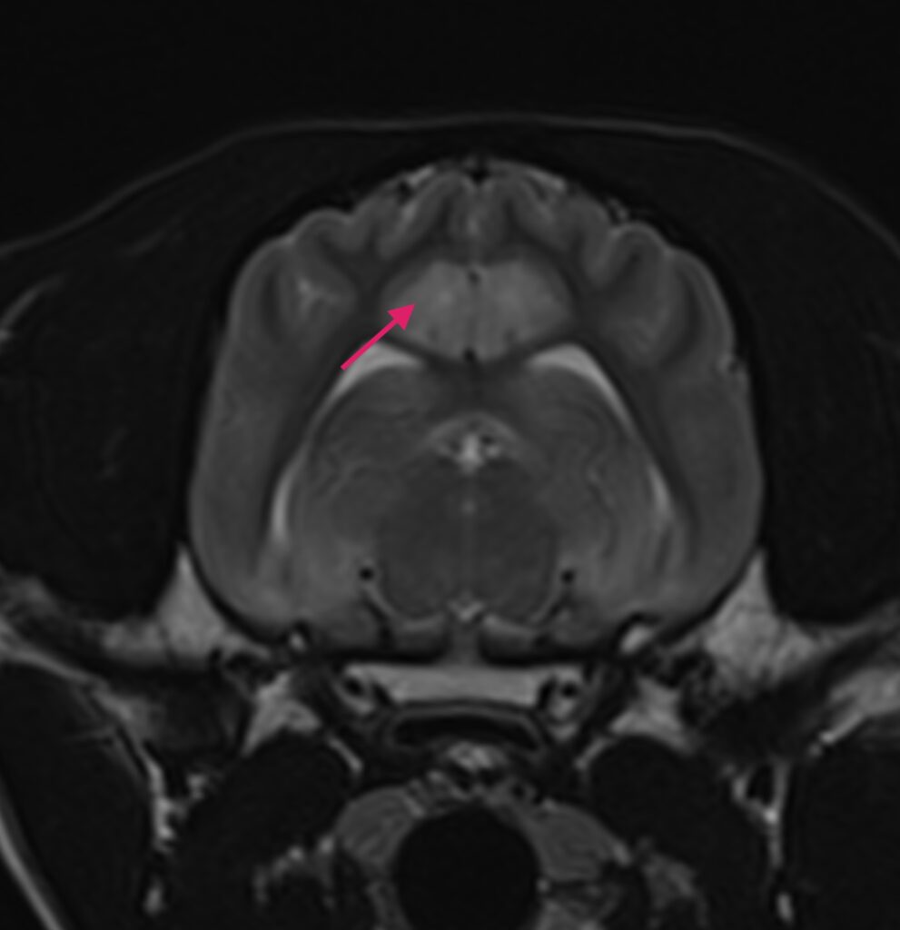
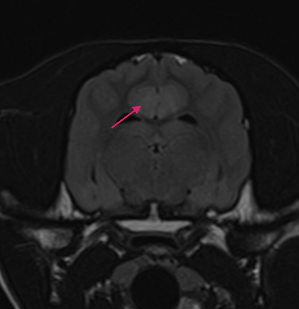
All lesions are T2 hyperintense without suppression on FLAIR sequences. T1 sequences show lesions to be iso to hypointense to the surrounding tissue. The vast majority of the lesions are bilateral and symmetrical. The distribution patterns support existing ideas of connectivity through the above regions.


Figure 1A – a transverse T2W (A) and FLAIR (B) image of a 1-year-old Labrador retriever 2 days after experiencing cluster seizures. There is hyperintensity of the cingulate gyri (arrows) on both images.
A Similarity in People and Pets?
Some of the more common links are between the cingulate gyrus and parahippocampal gyrus, the cingulate gyrus and pulvinar thalamic nucleus as well as the hippocampus and parahippocampus. These locations and connections are similar to what has been reported in the human seizure activity, particularly the pulvinar thalamic nucleus which is important to the genesis and reciprocal propagation of the seizures.
Conclusion
It is clear that more work and evaluation are needed to better understand the pathogenesis of the post-ictal changes and the client observed manifestations of these changes. With this knowledge, we will be able to complete the circle of treatment from epileptic seizure through the post-ictal and recovery phases.
References
[1] Kähn C, Meyerhoff N, Meller S, Nessler JN, Volk HA, Charalambous M. The Postictal Phase in Canine Idiopathic Epilepsy: Semiology, Management, and Impact on the Quality of Life from the Owners’ Perspective. Animals. 2024; 14(1):103. https://doi.org/10.3390/ani14010103Photo by Fermin Rodriguez Penelas on Unsplash


