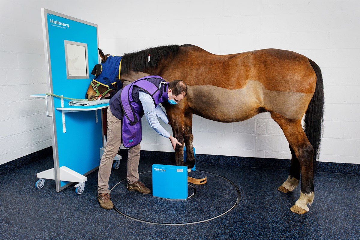
In this case study, kindly provided by Donnington Grove Equine Vets, we take a look at the case of a 6 year-old-gelding who was admitted for further diagnostic imaging of a left hindlimb lesion noted on radiographs. The horse was not lame.
Radiograph: the findings
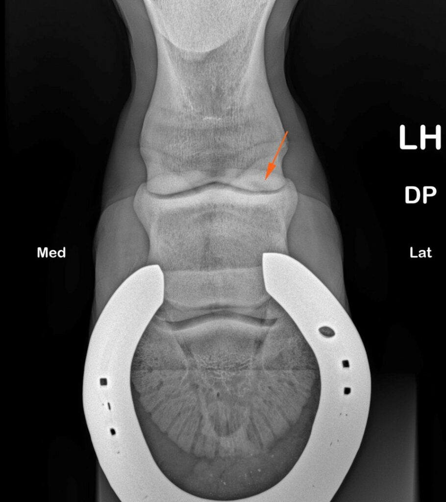
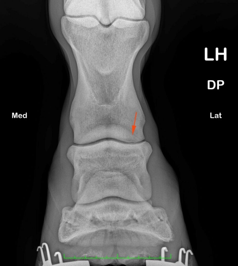
Figures one and two show lucency in the lateral condyle of the proximal phalanx noted on radiographs (orange arrows).
CT: the findings
Imaging with Hallmarq’s Vision CT enabled a more quantitative assessment and revealed an elongated defect of the subchondral bone plate, at the lateral aspect of the pastern joint, involving the proximal and middle phalanges. Within the distal subchondral bone plate of the proximal phalanx, it presented as a shallow elongated concave defect measuring approximately 3mm in mediolateral width and 17mm in dorsoplantar length, extending in a dorsolateral to plantaromedial direction (green arrows).
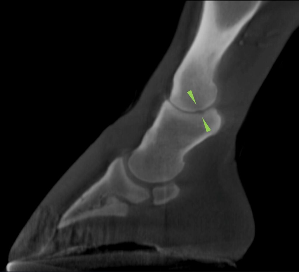
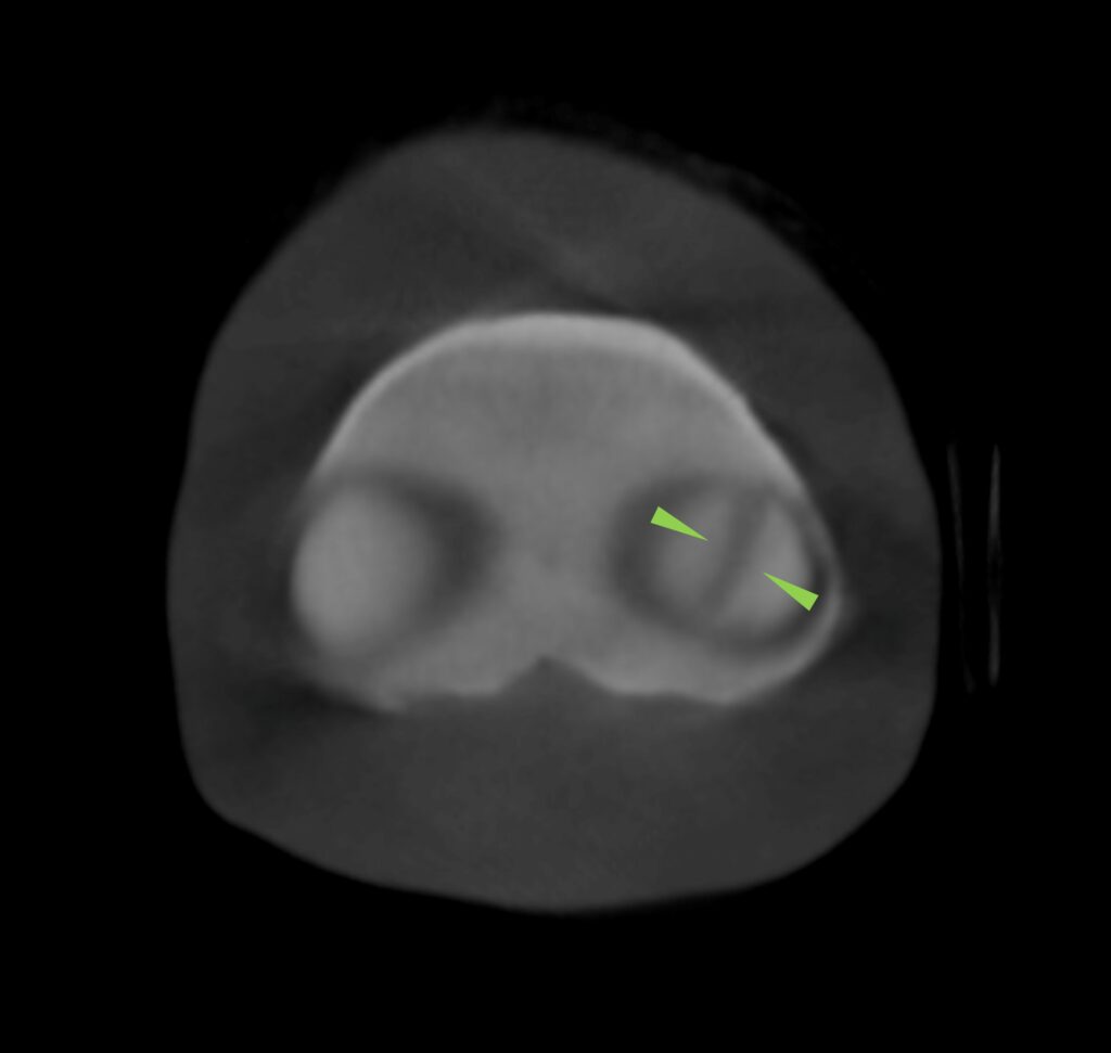
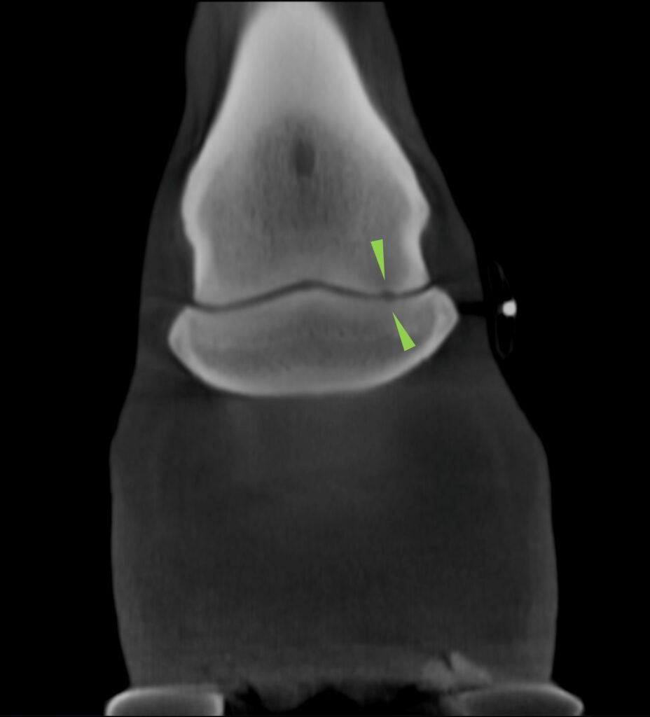
Figures 3, 4, and 5 show that a small and poorly marginated region of hypoattenuation of the adjacent trabecular bone is also present. A shallower kissing lesion extends in the same direction through the subchondral bone plate of the lateral glenoid of the middle phalanx with no extension to the adjacent trabecular bone.
Prognosis
It was considered that this unusual lesion possibly represented a small OCD-type lesion, not unlike the ‘dimple’ lesions seen in stifles, representing a small and less significant version of a bone cyst. The lesion is most likely to have been present for many years and at present there is no evidence of associated osteoarthritis. In light of the soundness of the horse, currently, the lesion would be best monitored.
With thanks to Donnington Grove Equine Vets, UK, for sharing this case with us.