Thoroughbred gelding with right forelimb lameness
In this case study, kindly provided by Donnington Grove Equine Vets, we take a look at the case of a 2-year-old thoroughbred gelding, admitted with a one-week history of right forelimb lameness.
History
Exploration of the sole and radiographic examination was unremarkable. A tract could not be visualised and the patient was admitted to the clinic with the suspicion of a solar abscess.
MRI findings
MR imaging of the foot revealed a chronic penetrating tract, severe infectious osteitis of the pedal bone, and osseous changes at the solar canal.
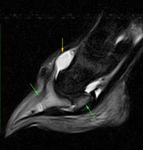
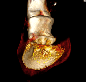
Figure 1 (above) – MRI image showing diffuse bone oedema in the distal phalanx and navicular bone (green arrows) and effusion of the coffin joint (yellow arrow)
Standing surgery was performed in an attempt to widen the penetrating tract likely caused by a foreign body and allow better drainage and improve the horse’s comfort level. The patient’s comfort level did not improve over the next few days so further imaging, CT scan, was performed to allow quick reassessment of the tract and pedal bone.
CT findings
CT imaging showed the extent of the solar hoof defect and the extension of pathology both dorsally and proximally within the pedal bone.
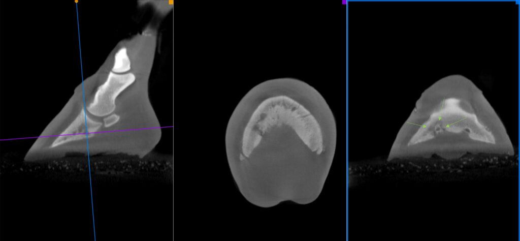
Figures 2, 2(a) and 2(b) showing the extent of osseous change around the solar canal (green arrows).
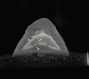
Subsequently, more aggressive debridement of the affected tissue within the pedal bone was undertaken under general anaesthesia. Repeat CT was performed 10 days after surgery to re-assess the pedal bone and ensure no further debridement was indicated.
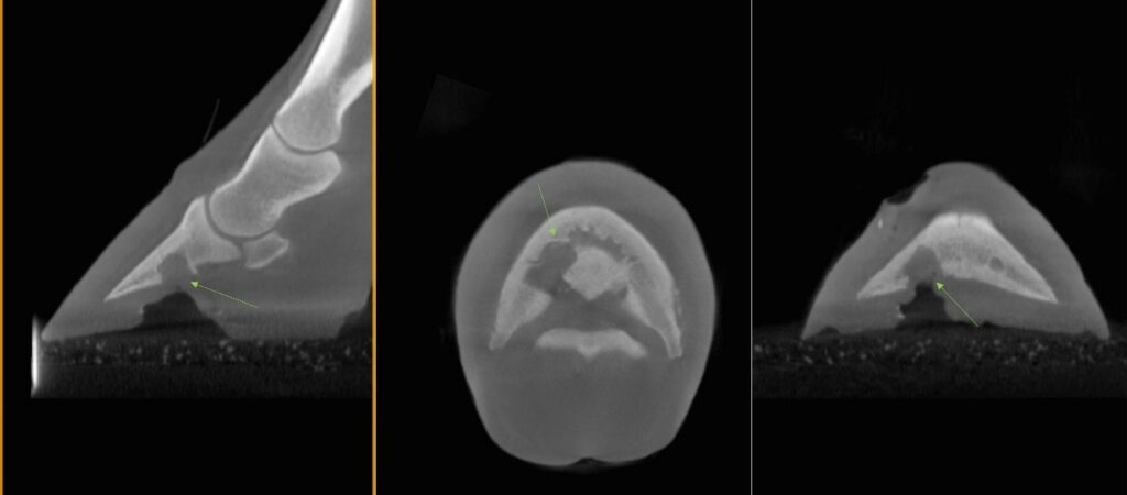
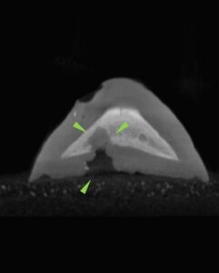
Figure 3, 3(a), and 3(b) show the repeat CT scan 10 days after surgery. The extent of pedal bone debrided is shown by green arrows.
Prognosis
The initial prognosis for this case was very guarded due to the extent of the infection. Happily, the patient was discharged to continue recovery and rehabilitation at home.
With thanks to surgeon Dr Henry O’Neill, Donnington Grove Equine Vets, UK, for sharing this case with us.