


Mare Presenting 3/5 Right Forelimb Lameness
History
In a case study, kindly provided by Pferdeklinik Seeburg, we examine how MRI and CT were used to diagnose acute lameness in a 4.5yr old Trakehner mare. The mare was 3/5 right forelimb lame at a trot in a straight line.
Pain was exacerbated when walking in a circle. In addition, an increase in lameness was elicited on soft ground when the horse was lunged. The mare was right forelimb flexion positive.
Clinical Examination
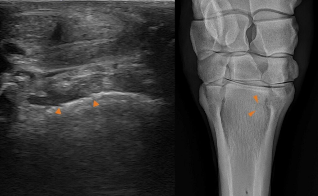
A positive response to lateral palmar nerve block, ultrasonographic and radiographic findings suggest suspensory origin pathology. Therefore, no further diagnostic analgesia was undertaken. Additionally, standing low field MRI was performed followed by CT for further interest.
MRI Findings
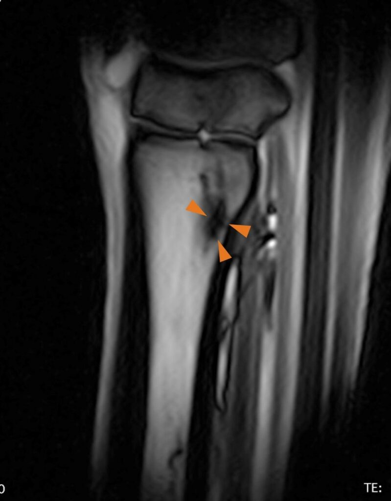
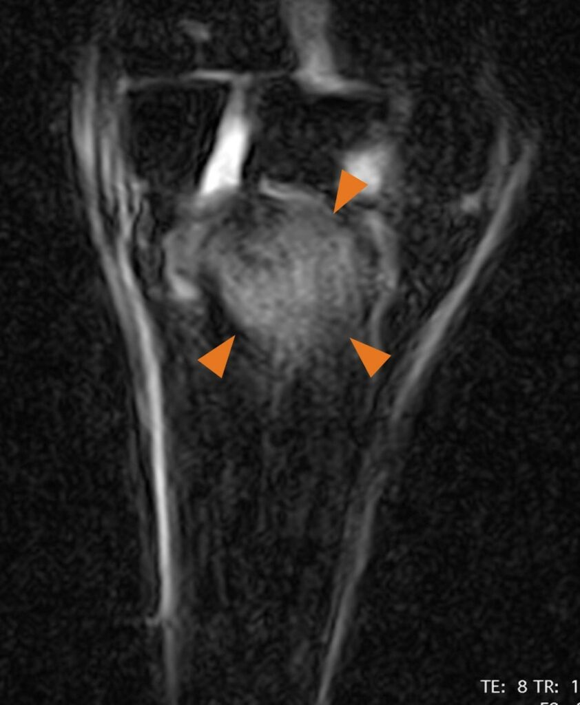
MR imaging illustrates signal increase in proximal palmar MCIII at the level of the suspensory origin. This is consistent with a high grade of bone edema at the origin of the suspensory ligament. It is not possible to visualize a bone fragment. However, in combination with x-ray, ultrasound findings and the severity of the other MR findings, a bone avulsion like lesion can be assumed.
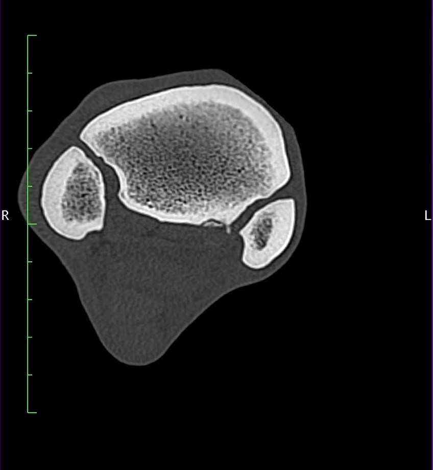
CT Findings
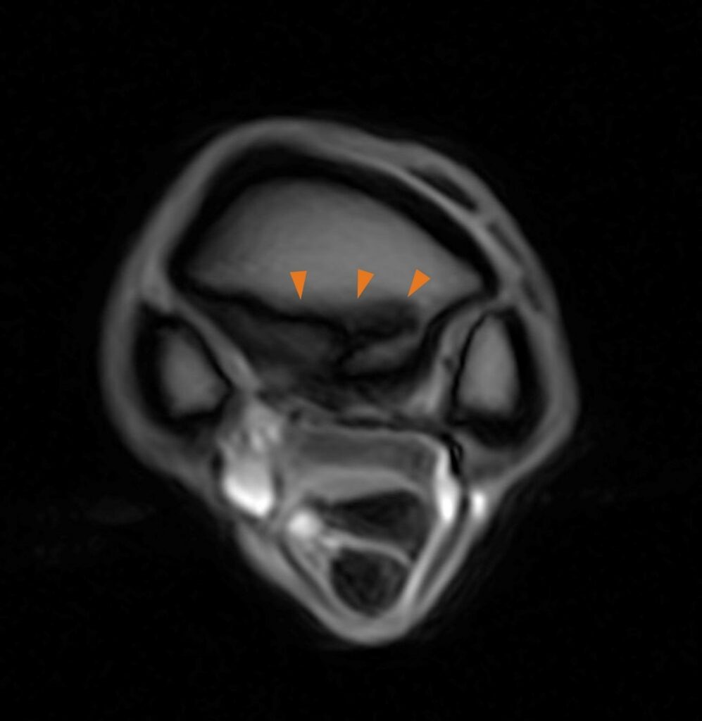
Avulsion fragment is clearly visible on the palmaro-medially proximal MCIII.
Diagnosis
In this case, proximal suspensory ligament desmopathy and enthesopathy associated with an avulsion fracture.
With thanks to Pferdeklinik Seeburg for providing this case study.