Looks like you’re on our UK site! Would you like to switch US site?

Do not ask again
Equine MRI

Introduction

MRI is within reach

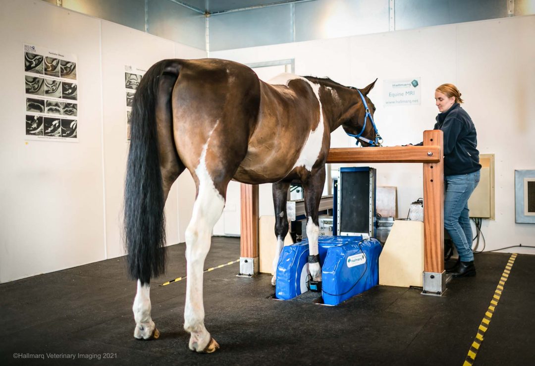
Standing Equine MRI (sMRI) is available for your practice – for everyday imaging cases 

  • Offer the gold standard for imaging quickly, safely and conveniently
  • Diagnostic in over 90% of cases, Standing Equine MRI accurately identifies the specific cause of lameness.
  • Conveniently bring all the benefits of MRI to your practice without the hassle or risk of general anesthesia.
  • Backed by Hallmarq Q-Care, you can reach profitability with just 5 cases per month.

Providing clinicians better guidance for treatment

Benefits of sMRI

More Accurate Diagnosis

Comprehensive MRI diagnosis allows you to evaluate the integrity of all the structures within a region, helping you make a more informed decision for treatment.

Informed Prognosis

Understand the full extent of the injury based on the degree of pathology in order to determine how aggressively you approach treatment.

Quick & Affordable Answers

Stop the guess work. Utilizing MRI as ‘first line’ imaging gets you an answer quickly and affordably. Protect the owner’s investment by definitively ruling out costly conditions that may not be causing any signs of lameness.

Visualize Tissues & Densities

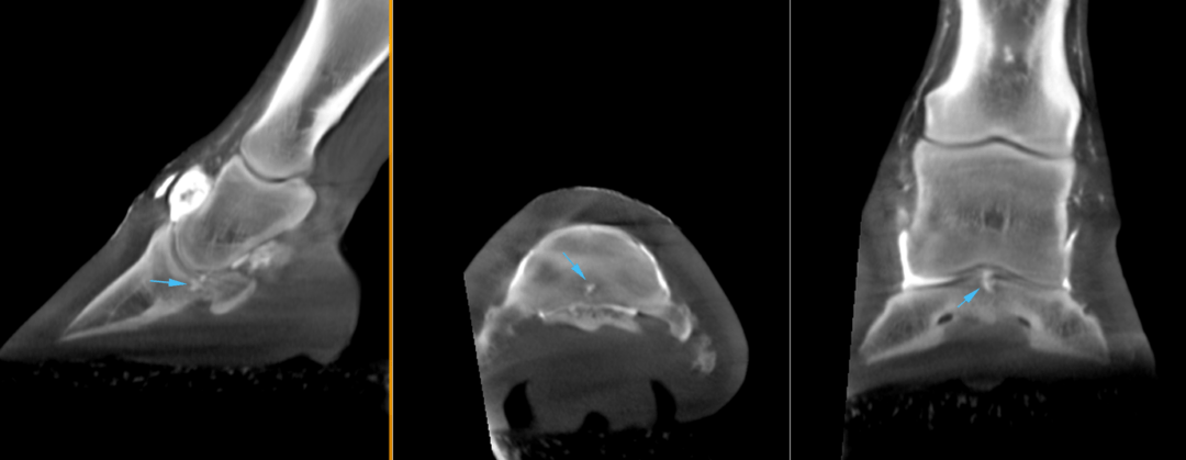
Far beyond radiography, ultrasonography and even CT in some cases, standing equine MRI is capable of assessing both bony and soft tissue injuries with a single procedure.

Effectively Guide Treatment

MRI is accessible to a wide range of clients and patients and works best as an initial or ‘first line’ imaging modality to guide treatment decisions from the beginning of therapy and rehabilitation.

A Clear Picture of Chronic Lameness

MRI gives a clear picture to see what was missing and preventing the horse from regaining complete soundness.

Hallmarq’s Standing Equine MRI (sMRI) system brings the same diagnostic capability to equine clinical practice as human MRI with the patient at the forefront of every design

Quick, Safe & Convenient MRI

Advantages of sMRI

MRI Without Anesthesia

Standing MRI allows the procedure to be more convenient and cost effective, while at the same time reducing the risk of potential complications of general anesthesia such as colic and poor recovery.

Scan Multiple Regions

Standing MRI can be performed on multiple regions of the distal limb including the tarsus, carpus, metacarpus/metatarsus, flexor sheath, annular ligament, MCP joint, pastern joint, flexor tendons, distal sesamoidean ligaments, collateral ligaments, coffin bone, DIP, impar ligament, navicular bone and more.

True Diagnostic Quality

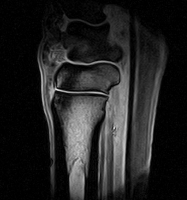
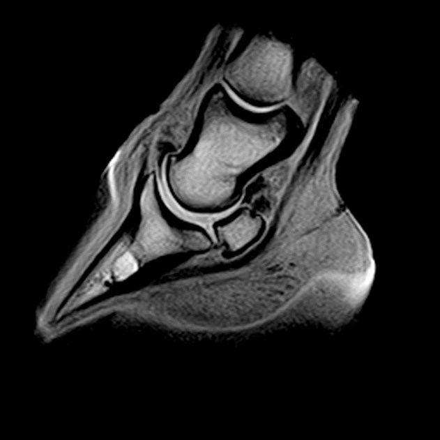
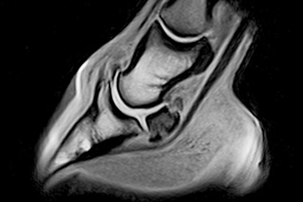
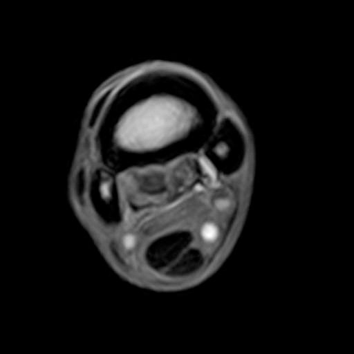
The diagnostic quality of standing (low-field) images has improved dramatically over the last 10 years. Standing equine MRI is now diagnostic in 90% of cases.

Radiologist Expertise

Radiologists are involved in the reading and reporting process whether remote or on-site. Radiologists have extensive experience with all types of MRI, both low- and high-field and are able to educate clinicians in development of scanning protocols to assure each anatomic region is appropriately imaged so nothing gets overlooked.

Convenience & Accessibility

With standing MRI, MRI no longer has to be reserved for advanced cases. Acute injuries where the lameness is localized to one region can benefit from an initial MRI to accurately guide therapy, saving costs on empirical treatments that may not be as successful as targeted treatments.

Offer the Gold Standard

The hassle of sending the horse hours away often prevents MRI from being a viable option. Investing in an MRI means investing in your client and patient base. Having MRI in-house allows you to not only offer your patients accurate diagnosis, but also positions you as a referral center.

Detailed Imaging

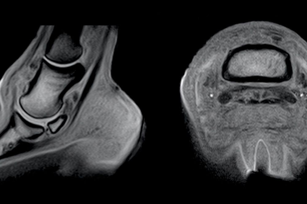
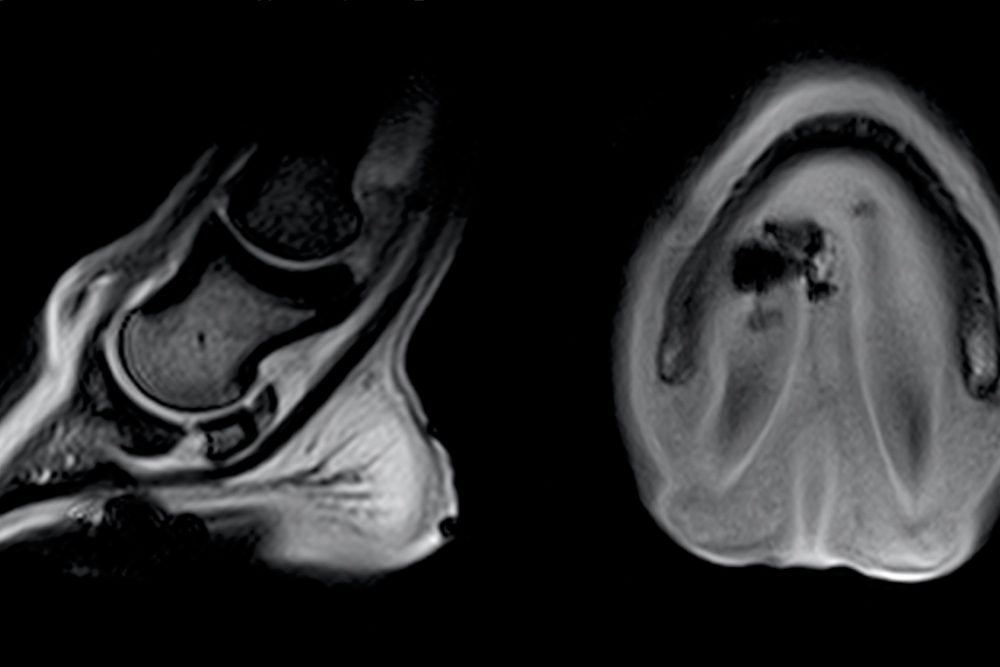
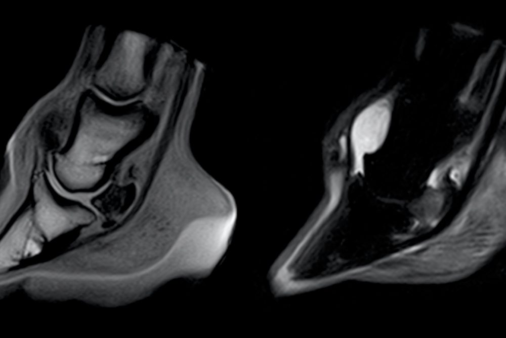
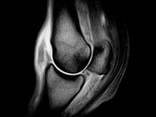
Clinical case gallery

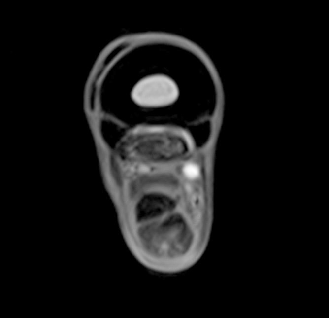
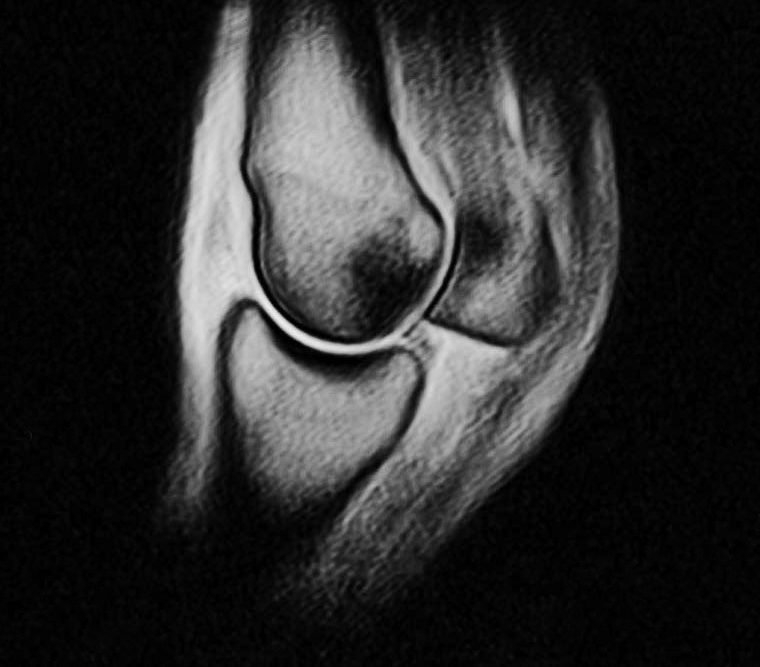
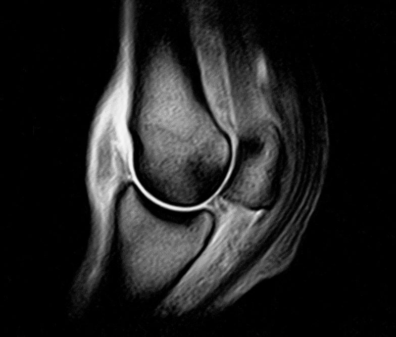
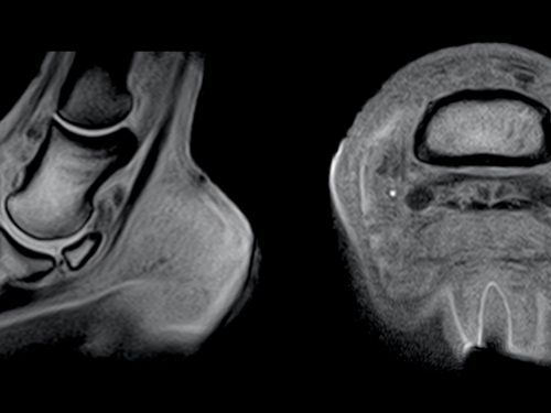
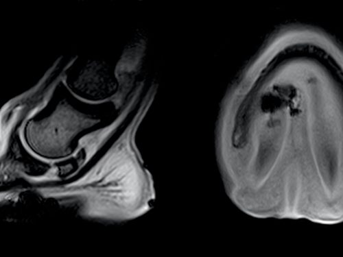
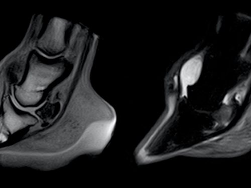
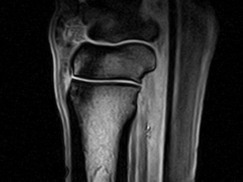
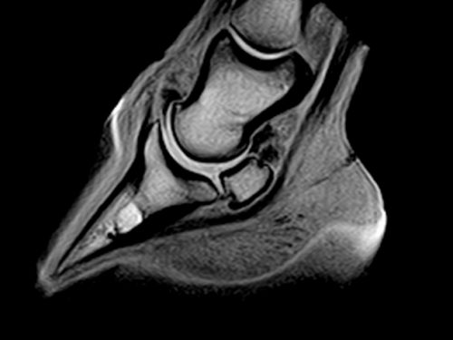
Specifically designed to image the limbs of standing horses, our innovative and award- winning equine MRI scanner captures superb images for a more accurate diagnosis. 

  • Excellent diagnostic images of the hoof, all the way up to the carpus/tarsus regions.
  • Patented design includes a unique open, thin poled magnet, bespoke equine optimized patient handling and equine specific RF coils.
  • Award-winning motion correction software, to ensure accurate diagnosis of the standing horse under only mild sedation.
  • Veterinary specific software including an array of MRI scan options with equine optimized imaging protocols for efficiency and ease of use.

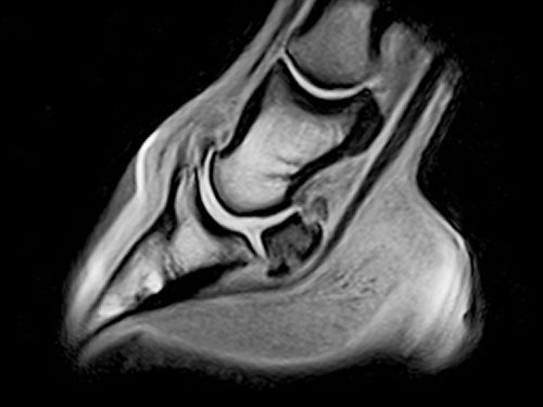
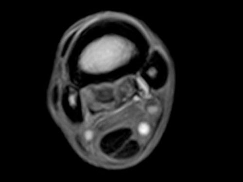
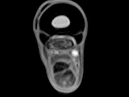
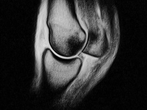
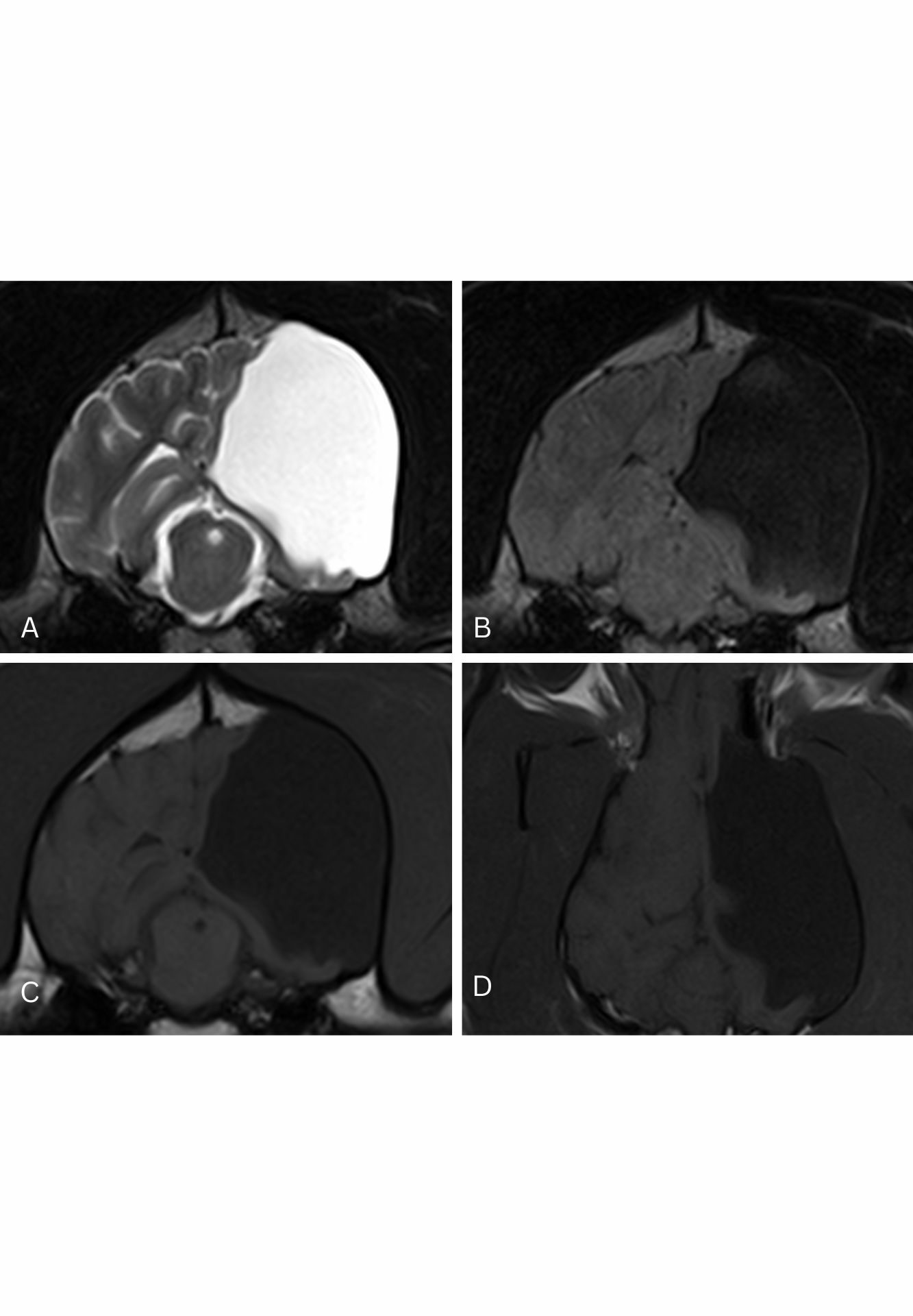
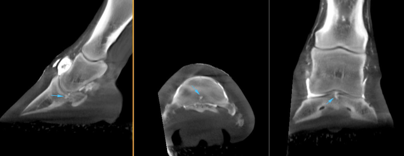
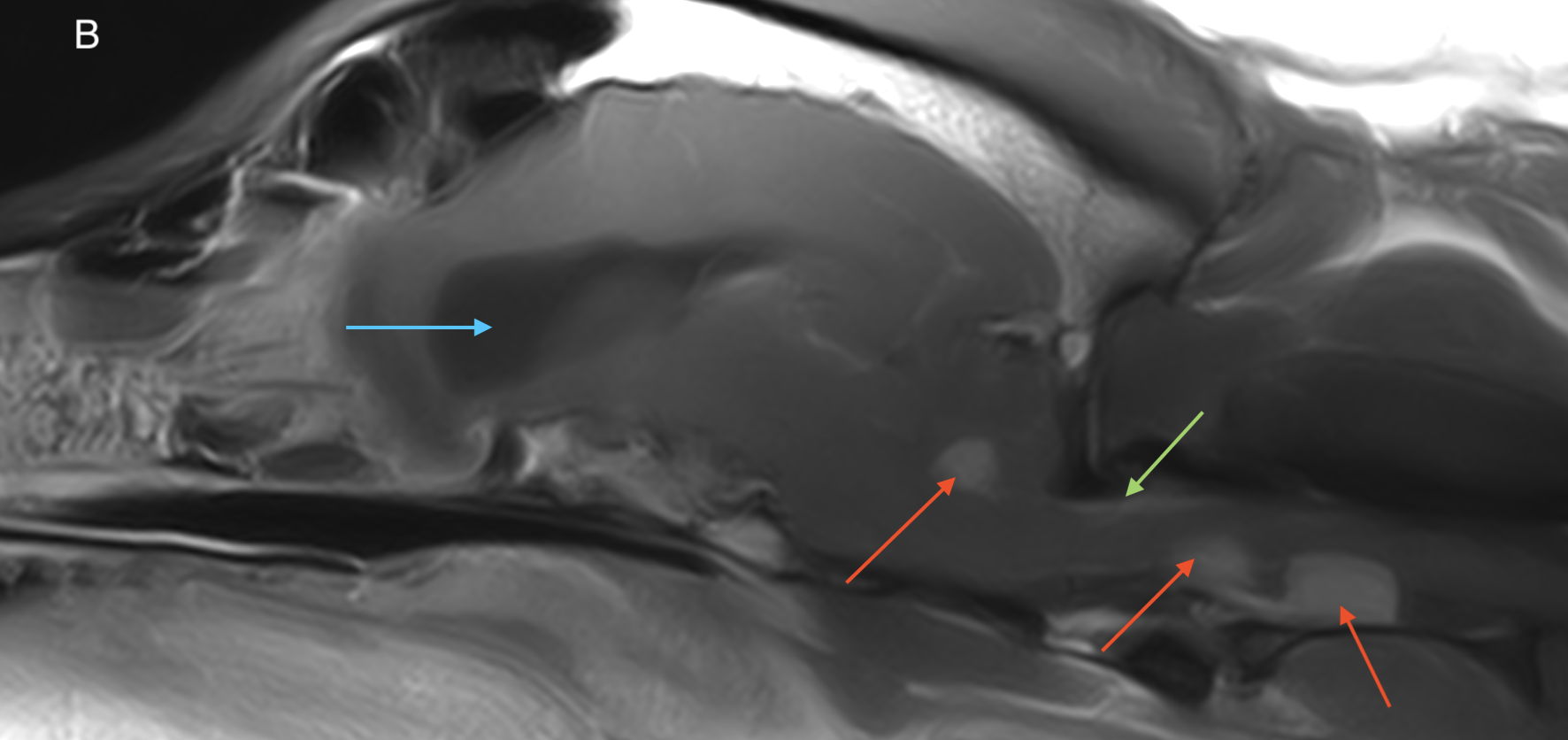
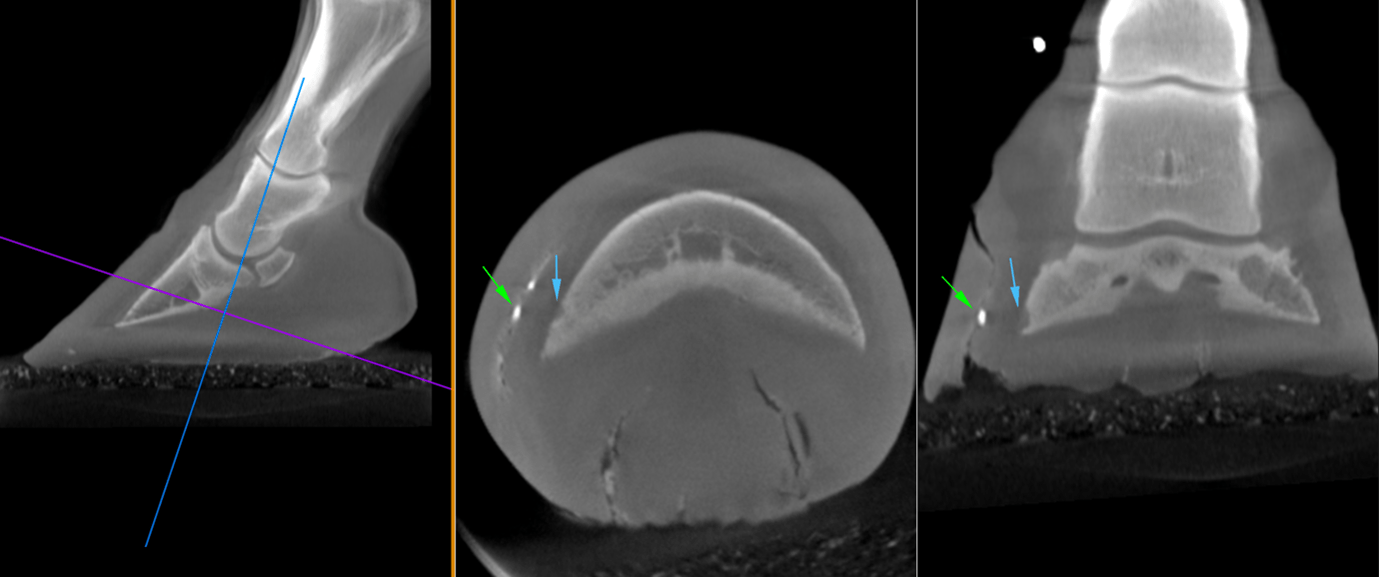
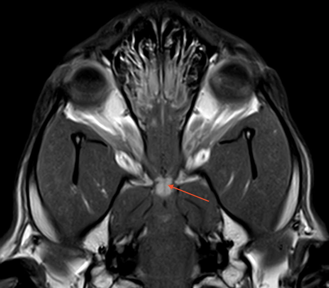
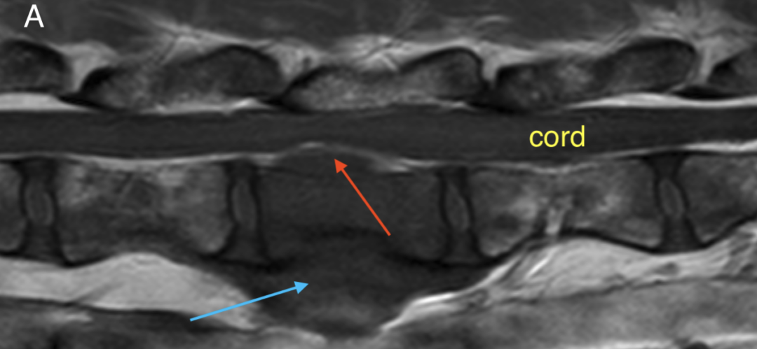
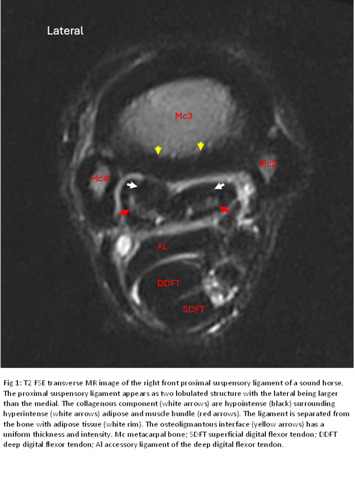
For real clinical examples from some of our installed scanners, please click to browse… 

Calculator

Hallmarq Standing Equine MRI is Affordable

With low upfront costs, built in seasonality and low running costs.

Low upfront costs

There are very low upfront costs – including an option to choose a modular solution instead of building out a room in the clinic.

Pay-as-you-go

You “pay-as-you-go”. Seasonality built in!

Profitable MRI service

With low running costs you can run a profitable MRI service with just a handful of cases per month. We have a unique financial model that reduces your investment risk.

Loading Equine MRI profit calculator…

Affordable, accessible, quick and convenient MRI

The process

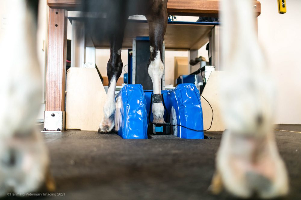
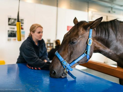
A vet with a horse in an MRI scanning room

Preparation

  • General physical and lameness examination
  • Removal of shoes (for imaging feet) on affected limb and contralateral limb for comparison and to avoid artifact.
  • Radiographs – assure no nail clinches are left in the hoof that would create an artifact.
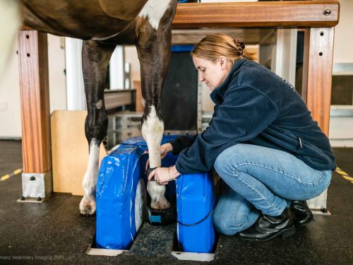
A vet preparing a horse for an MRI scanning

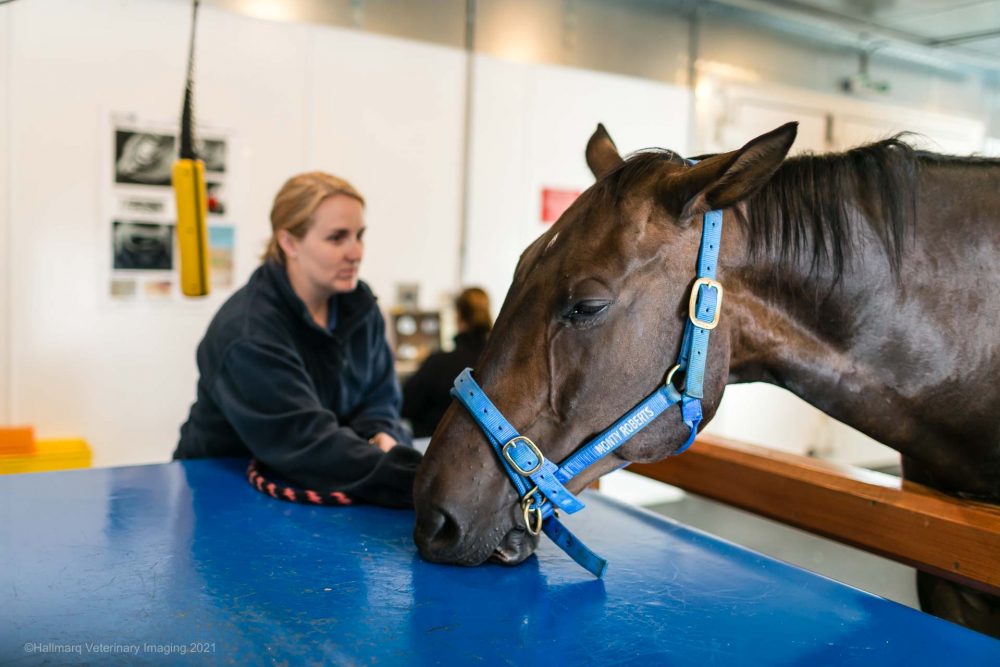
Sedation

  • Many protocols and procedure options to fit your clinic
  • Catheter or needle
  • CRI or “topped off” as necessary
  • Appropriate sedation typically achieved with alpha-2 agonists +/- opioid
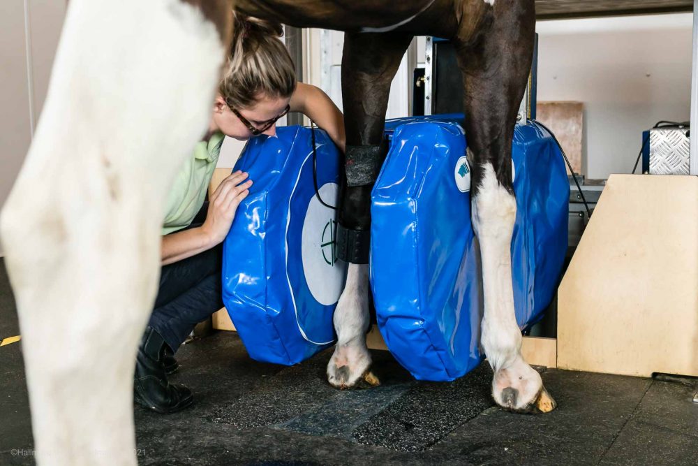
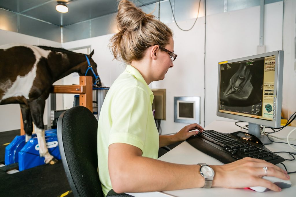
MRI operator, managing horse in Equine MRI scanner

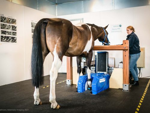
Positioning

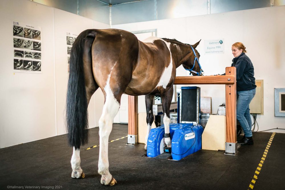
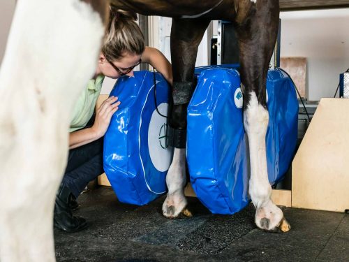
  • Sedated and walked into MRI
  • Lame leg usually positioned for scanning first
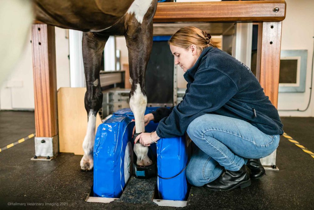
  • Radiofrequency coil placed around area of interest
  • Coil and magnet are both adjustable (within millimeters) to assure the region of interest is exactly in the middle of the scanning area of all 3 planes (x,y and z)
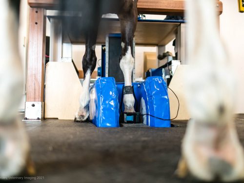
Equine MRI, standing MRI for Horses

Scanning

  • The first leg or region is scanned
  • A set of pilot images are acquired to assure region of interest is located in the center of the magnet
  • Multiple types of scans are set up in multiple planes to acquire all information about the region of interest
  • Second leg or region is scanned

Horse in Equine MRI scanner

Post Scan Recovery

  • Standing MRI makes outpatient visits possible
  • The horse is placed in a stall or cross-ties to recover from sedation
  • Generally, the horse can safely return home the same day
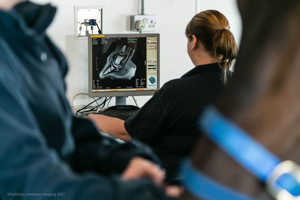
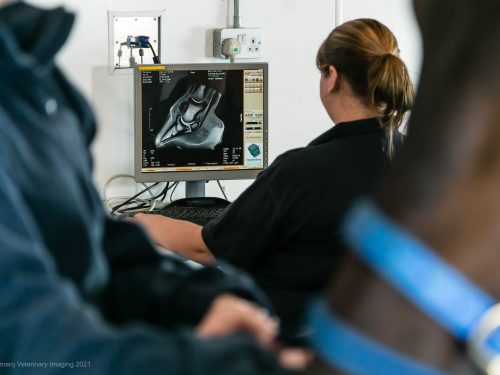
Vets reviewing an Equine MRI image

Diagnosis & Treatment

  • Interpretation – Radiologists are available for consultation and official report distribution (usually within 24 hours)
  • Consultation – Veterinarian and/or radiologist discuss the finding with the client
  • Treatment – With a conclusive diagnosis more targeted therapies are recommended and more accurate prognosis is given
  • Rehabilitation – Recommendations are provided to the client

Hallmarq MRI is convenient

Definitive diagnosis, better treatment and ultimate support.

Standing equine MRI can be used for providing diagnostic-quality images of soft tissue and boney structures in the distal limb. It’s not just for the foot!
The entire process is an outpatient procedure and does not require anesthesia. Offer a definitive diagnosis for a better treatment plan.
You are fully supported with the exclusive Q-Care world-class support program. With remote, onsite and online services, you are fully supported in building your MRI service.

“I must thank the whole Hallmarq team for your help and development of such a useful and effective tool”

Matteo Toniato, DVM Clinica Equina San Biagio
Hallmarq

“Standing MRI allows us to accurately diagnose the cause of lameness in the vast majority of cases where standard diagnostic techniques fail to give us the answer. It permits the selection of appropriate treatment methods, whereas without it we would often have been guessing.”

Tim Mair BVSc, PhD, DEIM, DESTS, DipECEIM, MRCVS Bell Equine Veterinary Hospital
Hallmarq

“In many lameness cases, it is not possible to find a definitive diagnosis. With our Hallmarq system we are finally able to solve the puzzle! This is a big step ahead for us, our clients and most importantly, the horses.”

Dr. Martin Stoeckli Pferdeklinik Dalchenhof, Switzerland

“The team at Hallmarq continue to find innovative approaches to improve motion correction and reduce slice thickness. When I use their images at International conferences, I am repeatedly asked if they were taken with a high field magnet.”

Sarah Taylor BVM&S MSc PhD Cert ES(Orth) DipECVS DipECVSMR MRCVS The University of Edinburgh. The Royal (Dick) School of Veterinary Studies
Hallmarq

“We are proud to have been involved with the development of Hallmarq’s standing MRI scanner since its inception. This technology has transformed our ability to diagnose many musculoskeletal disorders, particularly diseases affecting the foot.”

Tim Mair BVSc, PhD, DEIM, DESTS, DipECEIM, MRCVS Bell Equine Veterinary Hospital

“Hallmarq has been a great partner in providing our MRI services. The training provided was well organized and sponsorship of our MRI CE program provided to referring veterinarians was a great success. Thank you Hallmarq for your continued support.”

Dr. Bill Hay, DVM. DACVS Tryon Equine Hospital, Columbus, NC

“Hallmarq’s MRI has revolutionised the way foot lameness has been diagnosed in thousands of cases over the last 15 years. Excellent image quality facilitates accurate diagnoses, appropriate treatment selection and formulation of a prognosis.”

Sarah Taylor BVM&S MSc PhD Cert ES(Orth) DipECVS DipECVSMR MRCVS The University of Edinburgh. The Royal (Dick) School of Veterinary Studies

“The team at Hallmarq provide superb customer support, are friendly, approachable and always happy to help. This enables the user to make the most of the system and optimize image quality.”

Dr Rhiannon Morgan BSc BVSc CertAVP MVetMed(Hons) PhD DipECVDI MRCVS Royal Veterinary College

“Advanced diagnostic imaging techniques have become increasingly within reach for countless horses over the past 15 years and Hallmarq has played a large part in this rapid and exciting evolution with the equine veterinarian industry.”

Merry Smith MA VetMB PhD Cert ES DipECVS AECVDI MRCVS Image Equine

“Since installing Hallmarq’s Standing MRI in 2013, we have performed hundreds of examinations of the lower limbs, primarily in Thoroughbred racehorses. It is invaluable in identifying injuries that are not visible with conventional modalities.”

Dr Paul Robinson BVSc DACVS-LA The Hong Kong Jockey Club
Hallmarq

“We have used Hallmarq’s Standing Equine MRI for nearly 20 years. Hallmarq has consistently provided us with quality, leading-edge equipment, fantastic hands-on training for our staff, and excellent remote customer service.”

Dr Maureen Kelleher DVM DACVS Virginia Tech Marion DuPont Scott Equine Medical Center

Q-Care

We are your trusted partner for advanced veterinary imaging.

Providing high quality images is just the beginning. We’re here to help optimize your imaging service – from initial inquiry through installation and beyond. We guarantee support along your imaging journey, with an unrivalled program designed to add value to your practice.

Find out about Q-Care

FAQs

Why refer?

Much has been learned about the causes of equine lameness since the advent of MRI.  From the previously under-diagnosed, such as collateral desmitis of the distal interphalangeal joint, through the previously misunderstood, such as navicular disease, to the previously unknown, such as bone marrow edema, MRI has revolutionized our ability to provide a diagnosis and improve prognosis in equine lameness.

MRI is unparalleled in providing images of both soft and bony tissues.  Distinguishing water from fat, it highlights areas of pathology such as inflammation and bruising, in a way that radiography, CT, ultrasound or nuclear scintigraphy just can’t do.  By imaging the region of interest in slices orientated in any 3D plane, a lesion can be visualized without superimposition of adjacent structures.  Multiple views allow you to appreciate the full extent of the injury.
Details

Technical Specifications

In planning for the installation of your Hallmarq Standing Equine MRI system, the high-level technical specifications below should be taken into consideration. Please consult the Site Planning Guide for additional details.

Hardware & Software
Magnet
Magnet field strength0.27 Tesla
Gap20cm
Imaging volume14cm DSV
Position3-dimensional magnet positioning system
Rotation90-degree magnet rotation
Movement70cm vertical movement
ControlsManual
RF Coils3 x coils in varying sizes for foot and lower limb
Electronics
SpectrometerPentium computer
Digital RF unit
RF and gradient amplifiers
17” monitor
Interconnecting cabling set and connections to adjacent mains power and internet
Software
MRI Software Suite
Pulse Sequence Library
Equine Modular Room & Power
Standard Room & Power

References

References

Benefits of MRI

Denoix, J.‐M., Crevier, N., Roger, B. and Lebas, J.‐F. (1993), Magnetic resonance imaging of the equine foot. Veterinary Radiology & Ultrasound, 34: 405-411. doi:10.1111/j.1740-8261.1993.tb02029.x

Mair, T. S., Kinns, J., Jones, R. D., & Bolas, N. M. (2005). Magnetic resonance imaging of the distal limb of the standing horse. Equine Veterinary Education, 17(2), 74-78. doi: 10.1111/j.2042-3292.2005.tb00340.x

Murray R, Mair T. (2005) Use of magnetic resonance imaging in lameness diagnosis in the horse. In Practice, 27(3), 138-146. doi: 10.1136/inpract.27.3.138

Dyson, S.J., Murray, R. And Schramme, M.C. (2005), Lameness associated with foot pain: results of magnetic resonance imaging in 199 horses (January 2001‐December 2003) and response to treatment. Equine Veterinary Journal, 37: 113-121. doi:10.2746/0425164054223804

Dyson, S., Murray, R., Schramme, M. And Branch, M. (2003), Magnetic resonance imaging of the equine foot: 15 horses. Equine Veterinary Journal, 35: 18-26. doi:10.2746/042516403775467531

Sampson, S.N., Schneider, R.K., Gavin, P.R., Ho, C.P., Tucker, R.L. And Charles, E.M. (2009), Magnetic resonance imaging findings in horses with recent onset navicular syndrome but without radiographic abnormalities. Veterinary Radiology & Ultrasound, 50: 339-346. doi:10.1111/j.1740-8261.2009.01547.x

Murray, R. C., Dyson, S., Branch, M., & Schramme, M. (2007). Validation of magnetic resonance imaging use in equine limbs. Clinical Techniques in Equine Practice, 6(1), 26-36. Doi: 10.1053/j.ctep.2006.11.003

Labens, R,  Schramme, MC,  Murray, RC,  Bolas, N.  Standing low‐field MRI of the equine proximal metacarpal/metatarsal region is considered useful for diagnosing primary bone pathology and makes a positive contribution to case management: A prospective survey study. Vet Radiol Ultrasound.  2020; 61: 197– 205. doi.org/10.1111/vru.12824

Swagemakers, J. H., Vente, C., & Mageed, M. (2016). Equine foot MRI: past, present and future. Journal of Veterinary Research and Animal Husbandry, 1(1), 104-5.

Resources

Downloads

Find out more about us and our products, download image samples and resources.

See all downloads
Image of files covers