Lateral Condylar and Lateral Proximal Sesamoid Fractures
In this study, kindly provided by Donnington Grove Equine Vets, we look at the case of a 2-year-old thoroughbred racehorse who presented acutely right hind lame. Initial radiographs revealed an incomplete minimally displaced lateral condylar fracture.
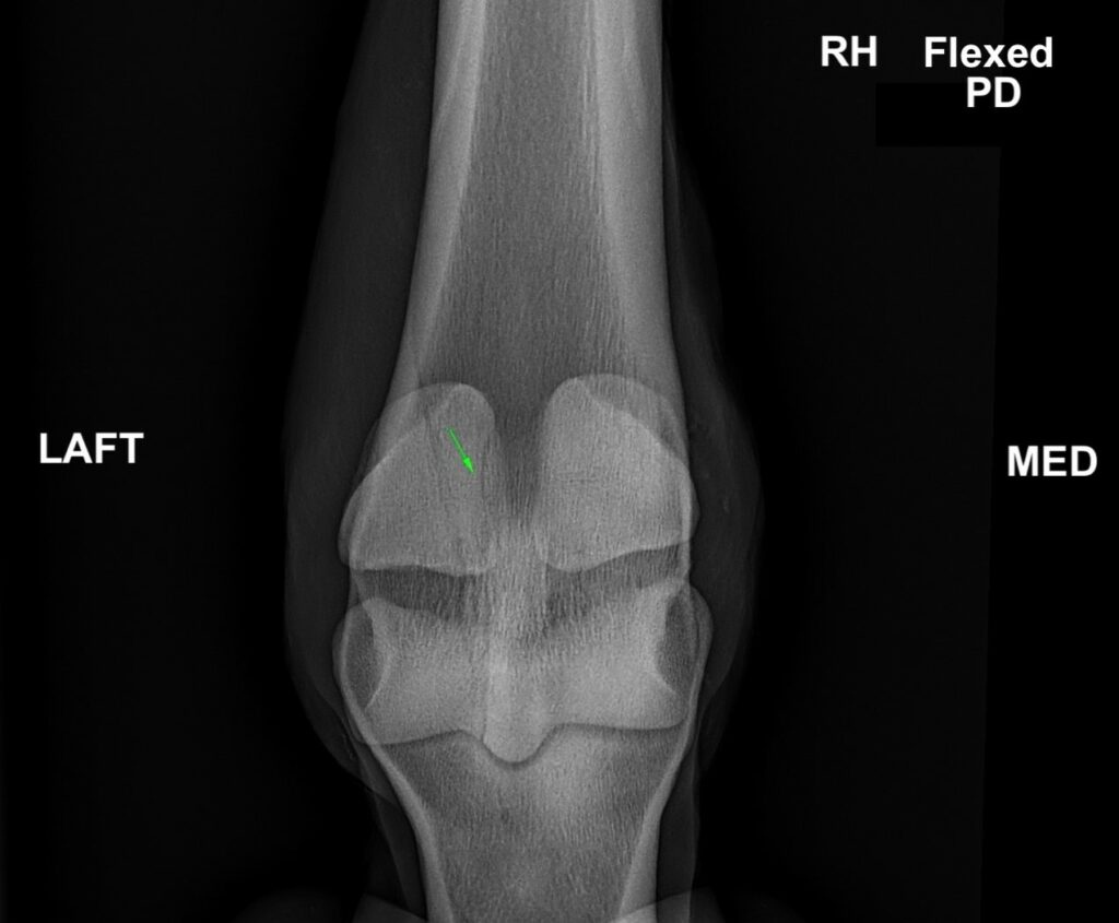
Radiographs
Upon admission to the hospital, additional radiographic projections were acquired. On flexed dorsoplantar radiographs, a radiolucent line was noted extending along the axial aspect of the lateral proximal sesamoid bone indicating a suspected additional fracture line.
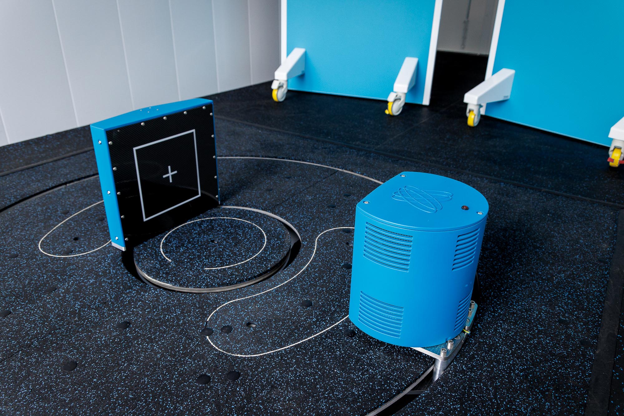
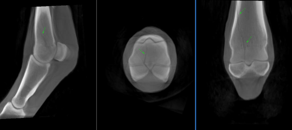
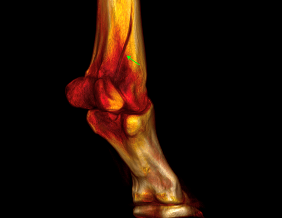
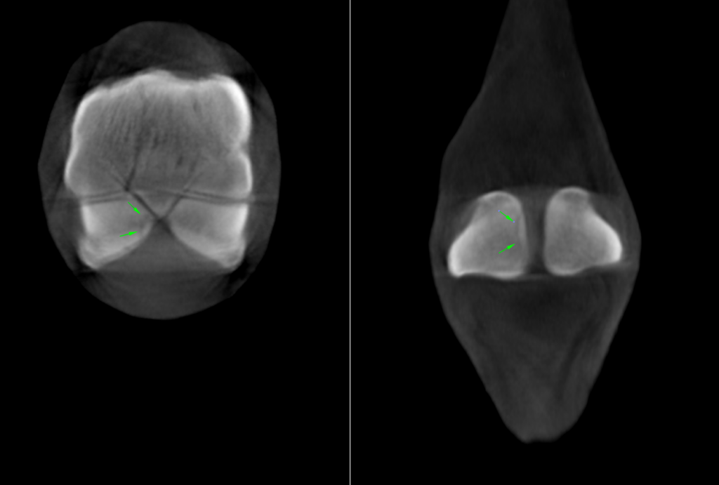
CT Findings
Further imaging with Hallmarq’s Vision CT enabled a more quantitative and precise evaluation, conclusively confirming the existence of a second non-displaced fracture line extending proximodistally through the axial aspect of the lateral proximal sesamoid bone.
The horse underwent surgical reduction and stabilization of the lateral condylar fracture under standing sedation. No additional treatment was pursued for the sesamoid fracture due to the challenges associated with accessing the site.
Prognosis
Although the treatment plan remained unchanged, the presence of the lateral sesamoid fracture impacted the prognosis, making it less favorable. The CT scan facilitated a more precise prognosis and informed adjustments to the management plan accordingly.
With thanks to surgeon Dr Henry O’Neill, Donnington Grove Equine Vets, UK, for sharing this case with us.