Just like humans who regularly turn to Magnetic Resonance Imaging (MRI) in the care and assessment of soft-tissue and bony injury, horses are frequently referred for what is considered the gold standard for diagnosing pathology associated with lameness.
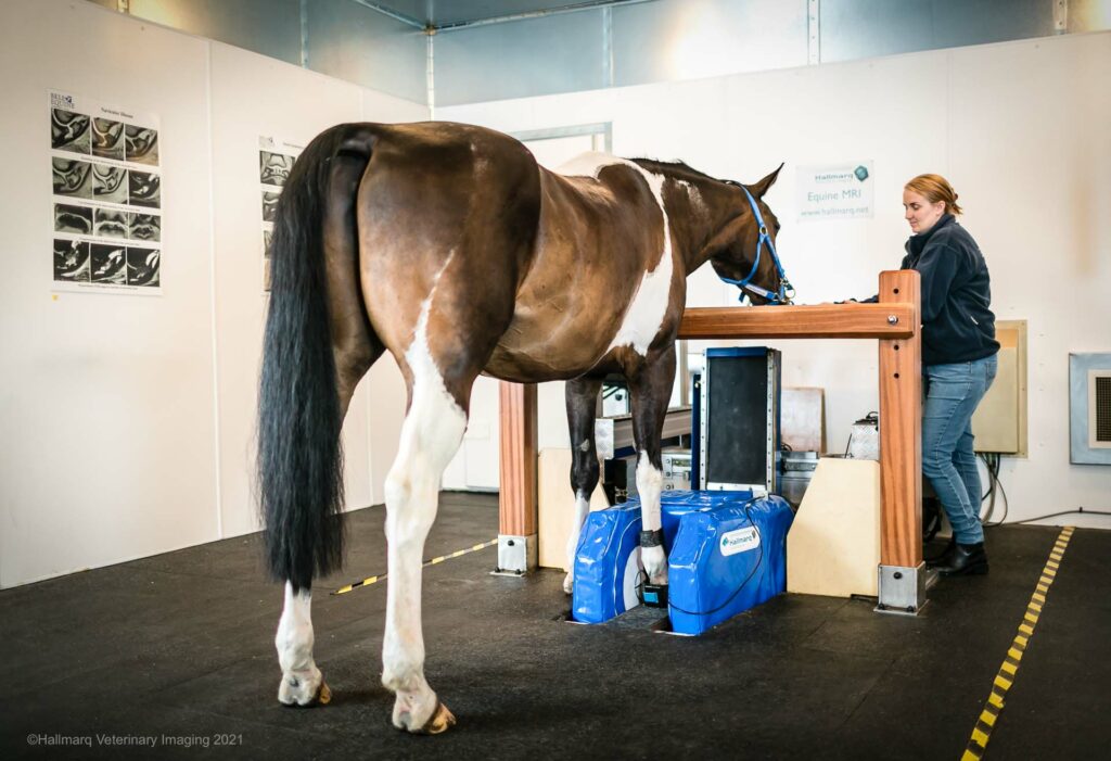
Machines optimized for human anatomy don’t lend themselves well to use in equine diagnostics which is why Hallmarq Veterinary Imaging pioneered their innovative Standing Equine MRI (sMRI) machine, designed specifically for the standing sedated horse. The alternative option is recumbent or ‘down’ MRI which requires the horse to be anesthetized
While both are valuable options for diagnosis, there are significant differences between the two when it comes to image quality, potential risks to the patient, and logistical requirements. Each has its pros and cons – here’s what you need to know.
Image Quality
Traditionally, down MRI, which often uses a high-field magnet, has delivered superior image quality with large tesla machines generally producing a more detailed image allowing for optimum diagnosis.
This, however, may no longer be the case. sMRI, which uses a low-field magnet, is now considered a viable alternative to rival its high-field alternatives, particularly when scanning the foot [2].
Hallmarq’s innovative technology has greatly advanced the quality of scans taken in the standing sedated horse. Accounting for a horse’s natural movement, and how that can impact image quality, their award-winning motion correction software automatically compensates for patient sway to produce images now thought comparable to its high-field counterparts
“The team at Hallmarq continues to find innovative approaches to improve motion correction and reduce slice thickness. When I use their images at international conferences, I am repeatedly asked if they were taken with a high-field machine.”
Sarah Taylor, BVM&S PhD CertES (Ortho) Dip ECVS DipECVSMR MRCVS The University of Edinburgh, The Royal (Dick) School of Veterinary Studies
What’s the Risk?
Down MRI requires the horse to undergo general anesthesia (GA) before image acquisition. Once anesthetized, the recumbent horse is positioned for scanning, with the distal limbs placed into the machine’s tubular core.
Despite the proficiency of practice staff in administering anesthesia and obtaining detailed images, many owners remain skeptical regarding the potential risks associated with down MRI. This is particularly the case for the already compromised horse not just at the time of scanning but in the weeks following.
About one in 100 healthy horses [1] is likely to undergo a post-anesthetic problem with the patient requiring close observation following the imaging procedure. For owners and trainers of elite sport horses, this risk is one that many are not willing to take; the risk versus benefit dilemma is one that only the horse owner can answer.
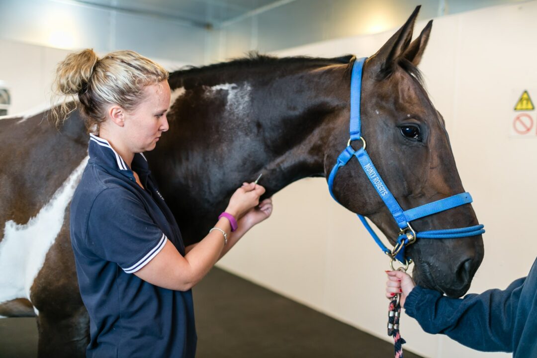
If the same information can be obtained without GA, however, then the question must be asked: why go for an elective imaging procedure? sMRI is risk-free, only requiring mild sedation for the acquisition of images thus providing a much safer option for the horse.
Cost Implications
Down MRI undoubtedly places a larger demand on practice staff with highly skilled personnel needed to administer anesthesia, and several people required to ensure the safe positioning of the horse in the magnet. The patient will need constant monitoring for the duration of the scan process. These additional costs are, of course, passed on to the horse owner.
In comparison, sMRI can be operated by just two staff – one qualified by Hallmarq to operate the machine and one to manage the patient. Since sMRI does not require GA, image acquisition is complete in a matter of hours as opposed to the lengthier procedure that down MRI demands. The practice and horse owner benefit from associated cost savings and most veterinarians can see more patients in less time, thus generating more revenue.
sMRI costs less to install, requires less space, and needs less shielding than high-field MRI. The stronger the magnet, the more care is needed in planning the surrounding area to minimize interference with image quality. Down MRI requires a large, dedicated room taking up valuable clinic space. In comparison, Hallmarq’s sMRI can be delivered in a self-contained modular room and dropped into place for quick and easy setup. The patient is simply walked into the magnet, a hoof coil attached to the relevant limb and scanning can start – a much less labor-intensive process than that required by down MRI.
What’s the Answer?
As with all decisions facing clinicians, there is no easy answer. All would agree that image quality is paramount, however, down and standing equine MRI have similar diagnostic capabilities. Both modalities generally result in the same diagnoses with diagnostic rates for distal limb lesions now comparable in both “up” and “down” MRI machines [3]. That advantage, when combined with potential cost savings and patient safety, surely makes standing a sensible solution.
[1] Dugdale A.H., Obhrai. J., Cripps P.J. (2016) Twenty years later: a single-centre, repeat retrospective analysis of equine perioperative mortality and investigation of recovery quality. Vet Anaesth Analg. 43, (2), 171-8
[2] Gutierrez-Nibeyro S, Werpy N, White Ii N. (2012) Standing low-field magnetic resonance imaging in horses with chronic foot pain. Aust Vet J. Mar, 90(3), pp 75-83
[3] Byrne, C.A., Marshall, J.F., Voute, L.C. (2020) Clinical magnetic resonance image quality of the equine foot is significantly influenced by acquisition system. Equine Vet J. 00, pp 1– 12
INTERESTED IN VISIONARY VETERINARY IMAGING?