Coffin Bone Fracture and Mild DDFT Lesion
Patient Information
This case study is kindly provided by Dr. Chris Bell, owner of Elders Equine Clinic, Manitoba. The case details a 12-year-old Quarter Horse gelding, imaged with Hallmarq’s Standing Equine MRI and Vision CT. With combination scanning using both modalities, a coffin bone fracture/fissure (type 2 fracture) and mild DDFT lesion in the medial lobe in the mid-pastern to about the top of the navicular bone margin was diagnosed.
Clinical History
The gelding has had a right front lameness, grade 3/5, for approximately 6 weeks. Blocks to abaxial with partial block to PD. Switches to left front lameness grade 1-2/5.
Radiographs show navicular sclerosis, some distal margin surface irregularities, and vascular channel/synovial evaginations but no obvious issue on the bone/joint for the lameness. The referring clinician took X-rays.
MRI Findings
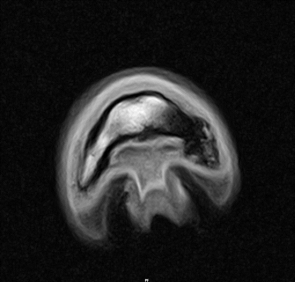
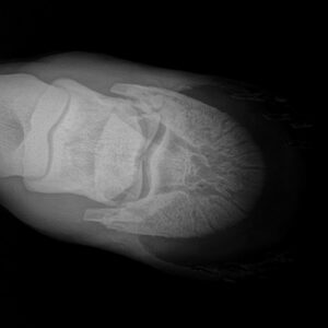
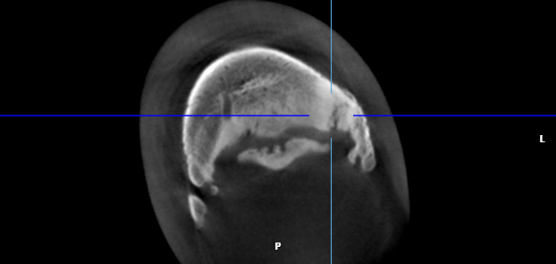
MRI was initiated and, during the exam, something in the coffin bone and the tissues surrounding the wing of the coffin bone and DDFT was apparent. This prompted a call to the owner and the suggestion that the horse be moved straight to CT following MRI.
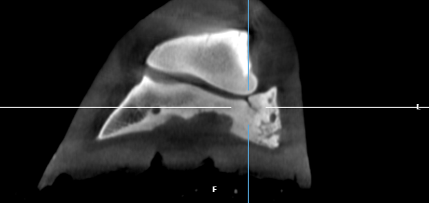
CT Findings
The fissure/fracture in the wing off the coffin bone was not appreciated or visible on the plain radiographs. However, it showed up clearly on the CT with MRI for supporting comparison of the extent of the fracture. The client appreciated the CT image of the fracture the best as a full 3D reconstruction was done on the premises and made available to her immediately.
“This was a very neat case. I’m glad the client decided to proceed to CT/MR, I don’t think we would have appreciated the extent of the fracture and soft tissue injuries without both modalities available.”
Dr. Chris Bell BSc, DVM, MVetSc, Board Certified Equine Surgeon (DACVS), Equine Surgery, Lameness and Sports Medicine, Elders Equine Veterinary Service
The Diagnosis
With combination scanning, a coffin bone fracture/fissure (type 2 fracture) and mild DDFT lesion in the medial lobe in the mid-pastern to about the top of the navicular bone margin was diagnosed.
The Recommendation
It was recommended to fit a bar shoe with quarter clips and float the medial heel with a recheck at 4 months. In addition, stall rest for 4-8 months depending on healing was advised. Recheck MRI or CT at 4-6 months. Prevequine 1 tablet once a day for 60 days. Ice the foot (optional) daily for 2 weeks.
A recheck MRI and CT will be performed to confirm fracture healing and whether consideration for surgical repair will be necessary.