Heel Bulb Laceration With Navicular Bone Fracture
In this case study, a 3-year-old thoroughbred gelding with a lateral heel bulb laceration of the left hind foot was referred for further investigation at Donnington Grove Equine Vets.
Radiographic Findings
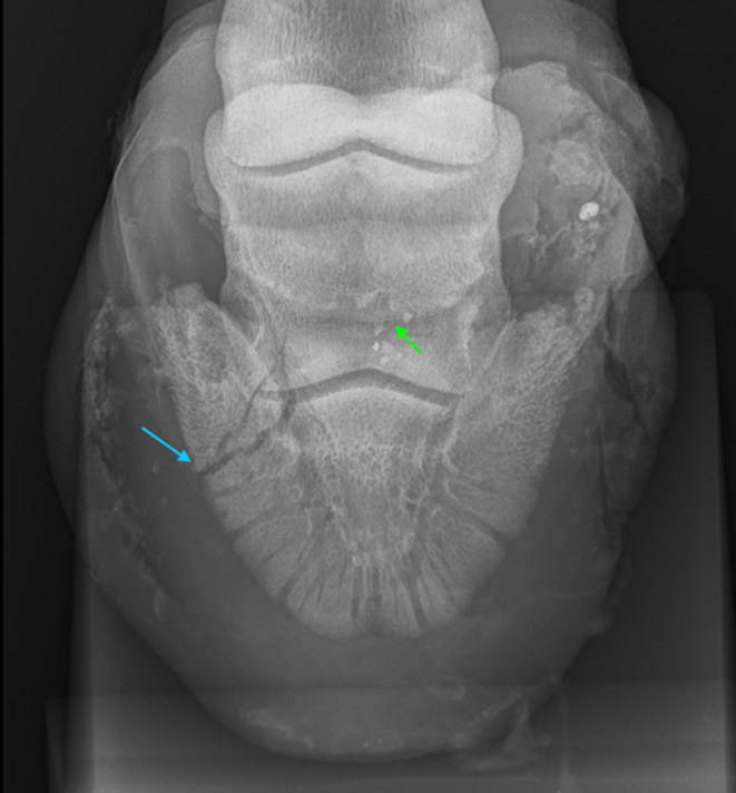
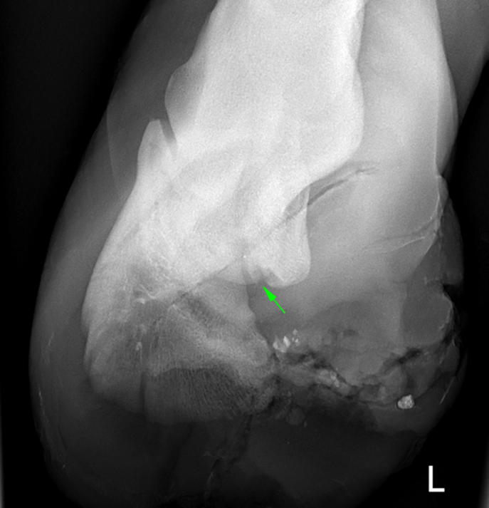
Radiographic examination of the foot was performed and identified a fracture of the medial wing of the distal phalanx (Figure 1 A).In addition, subtle irregularities were noted on the distal border of the navicular bone (Figure 1 B). Lateral to the right. The radiographs show the medial wing fracture (blue arrow) and the subtle irregularity of the navicular bone (green arrow).
CT Findings
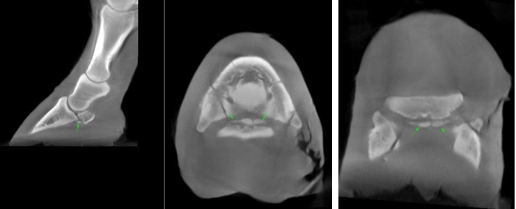
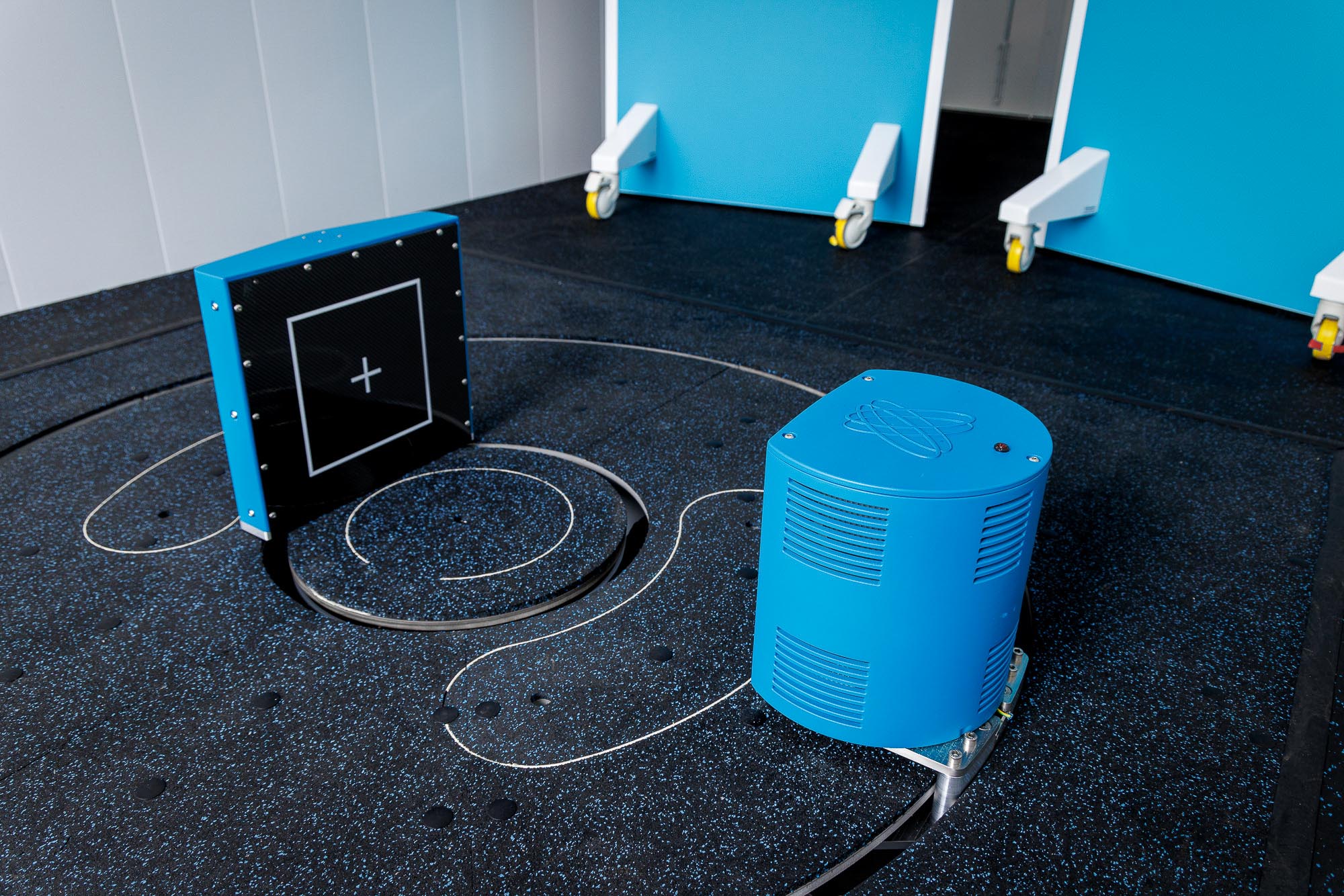
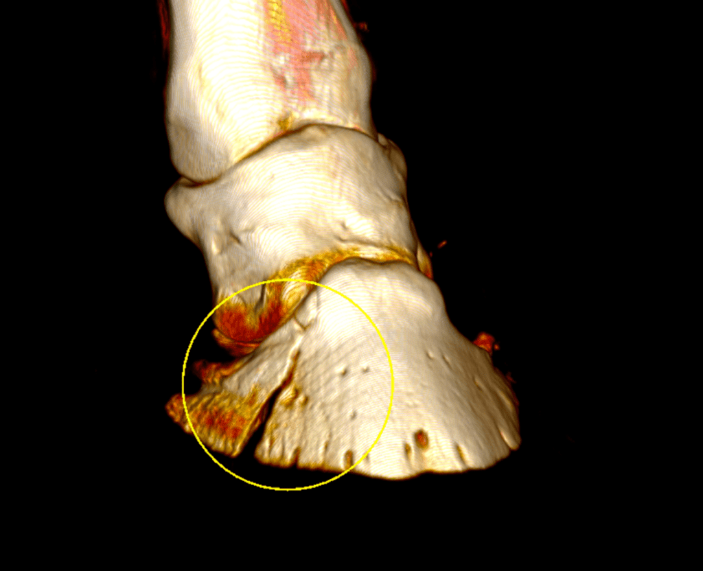
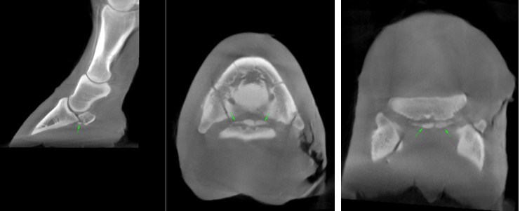
Scanning with Hallmarq’s Vision CT indicated an articular fracture of the medial wing of the distal phalanx. In addition, concurrent multiple fracture fragments were observed along the distal border of the navicular bone (Figures: 2,3).
Progression
Given the severity of the injuries, euthanasia was recommended. Post-mortem findings revealed that the wound at the heel bulb extended down to the lateral aspect of the navicular bursa.
Conclusion
CT can be superior to radiographs when assessing osseous changes in the navicular bone. Two-dimensional, radiographs can be limited by the superimposition of structures. In this case, Vision CT’s three-dimensional imaging provided a faster, more precise, and quantitative evaluation, clearly revealing the severity of the injury.
This case was kindly provided by Henry O’Neil MVB, DVM, MS, Dipl ACVS, MRCVS, Donnington Grove Equine Surgery, UK.
