


Intracranial Intra-arachnoid Diverticulum
The Patient
A 14-year-old male, neutered, Bichon Frise presented for the onset of seizure activity having exhibited 3 generalized tonic-clonic events over a 9-month period. A neurological examination revealed an absent left-sided menace response with a reduced right-sided response. However, no other neurological deficits were noted. Otherwise considered healthy, the dog underwent MRI using the Hallmarq 2nd generation 1.5T unit (see Fig 1).
The imaging results
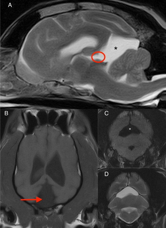
There is a large, sharply marginated, triangular-shaped cystic lesion in the caudo-dorsal cranial vault due to marked expansion of the quadrigeminal cistern (* and red arrow). This cystic lesion contains fluid isointense to CSF with no evidence of contrast uptake. It results in mild cranial and lateral displacement of the occipital lobes and in moderate caudoventral displacement and compression of the cerebellum. The thin membrane that normally separates the third ventricle from the quadrigeminal cistern is faintly visible on the sagittal image (red oval). The caudal margin of the cerebellum is flattened by the occipital bone and the caudal tip of the vermis is subtly impacting in foramen magnum as seen in (A). The rest of the brain study is unremarkable. The final diagnosis was an intracranial intra-arachnoid diverticulum.
(A) Sagittal T2W, (B) dorsal T1W, (C) transverse FLAIR and (D) transverse T2W images of the brain.
The most valuable MRI sequence for this case
The FLAIR image is important in this study to confirm that this fluid accumulation is compatible with CSF due to the suppression of signal seen, and not suggestive of a more highly proteinaceous accumulation of material which would incompletely suppress.
Disease overview
Intracranial intra-arachnoid cyst-like lesions represent accumulations of CSF that occur because of splitting or duplication of the arachnoid membrane. Because in some cases there is no evidence of a complete encapsulating membrane in which case the fluid is surrounded by normal tissue, the lesions are more appropriately termed diverticula (IAD). In dogs, these lesions are most frequently located within the caudal fossa; most seem to occur in a region comparable with the quadrigeminal cistern in humans and, therefore, have often been called quadrigeminal cysts.
Common Clinical Signs
Small breed brachycephalic male dogs seem to be predisposed to IADs with the most common breed reported being the Shih Tzu. In more than half of dogs retrospectively evaluated for intracranial disease, an IAD was considered an incidental finding. Clinical signs that are attributable to the cysts include focal or generalized seizures and cerebellar/vestibulocerebellar signs; however, paresis, a reduced level of consciousness, facial nerve paresis, and neck pain have also been reported. Overall, clinical signs reflect the location of the cyst and most likely occur as a result of compression of the neural tissue. Based on an MRI study, occipital lobe compression greater than 14% is likely to cause clinical signs, whereas the degree of cerebellar compression is not significantly associated with the occurrence of clinical signs. Seizures and headaches are the most common clinical signs in humans.
Typical MR imaging characteristics include (1) extra-axial, midline, frequently supracollicular location; (2) fluid similar in appearance to that of CSF; and (3) lack of contrast enhancement of the tissue immediately surrounding the fluid. A hypointense fluid-filled structure on T1-weighted images, which is hyperintense on T2- weighted images and fully suppresses with fluid-attenuated inversion recovery (FLAIR) imaging, is typically seen on MRI.
With thanks to IndyVet Emergency & Speciality Hospital, Indianapolis, IN, for providing this case study.
References
- Matiasek LA, Platt SR, Shaw S, Dennis R. Clinical and magnetic resonance imaging characteristics of quadrigeminal cysts in dogs. J Vet Intern Med. 2007 Sep-Oct;21(5):1021-6
- Platt S, Hicks J, Matiasek L. Intracranial Intra-arachnoid Diverticula and Cyst-like Abnormalities of the Brain. Vet Clin North Am Small Anim Pract. 2016 Mar;46(2):253-63
