

History
A 12-year-old Italian Saddlebred gelding was presented with an acute 3/5 left hindlimb lameness, more pronounced when trotted on the right rein on a soft surface. Clinical examination revealed a painful swelling over the plantar medial proximal metatarsal region. The lameness was abolished following a lateral plantar nerve block.
Ultrasonographic examination identified increased cross-sectional area of the lateral lobe of the proximal suspensory ligament. Radiographic examination was unremarkable.
MRI Findings
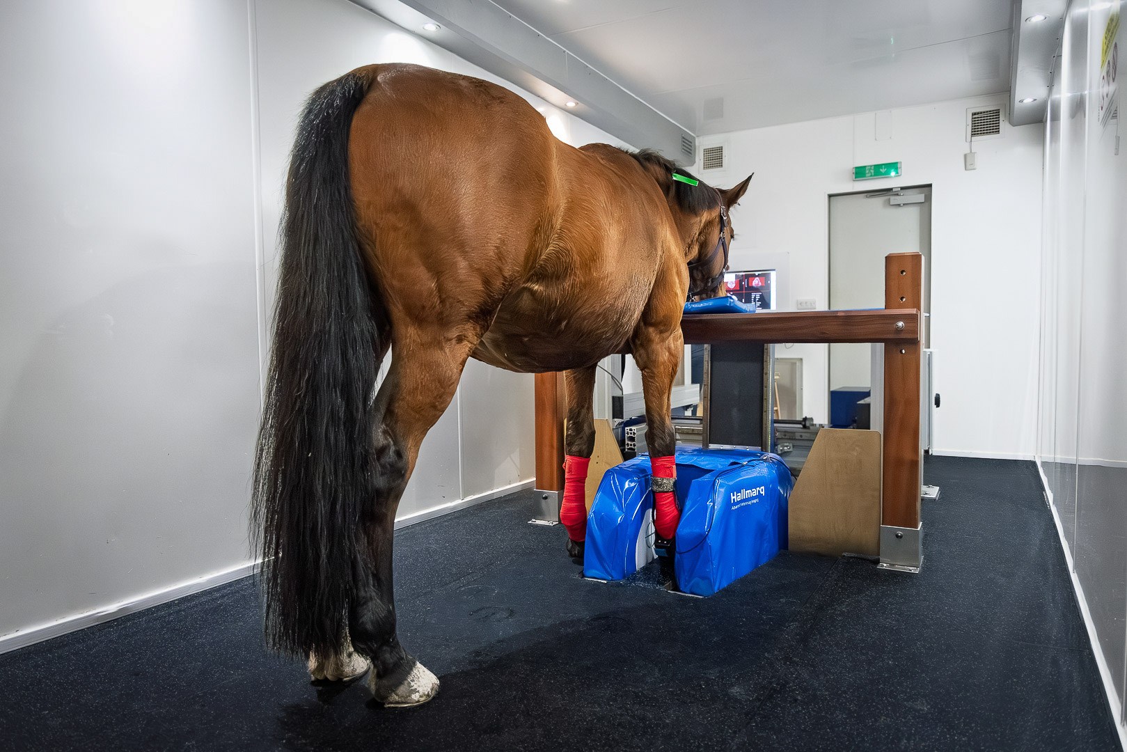
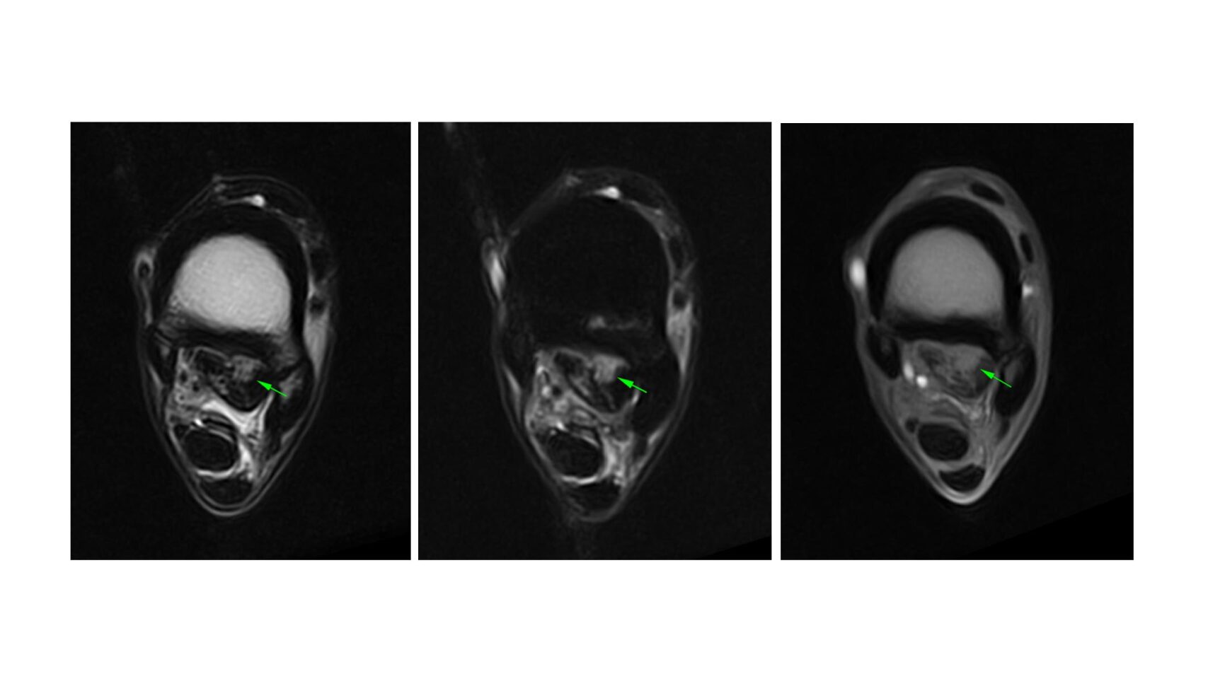
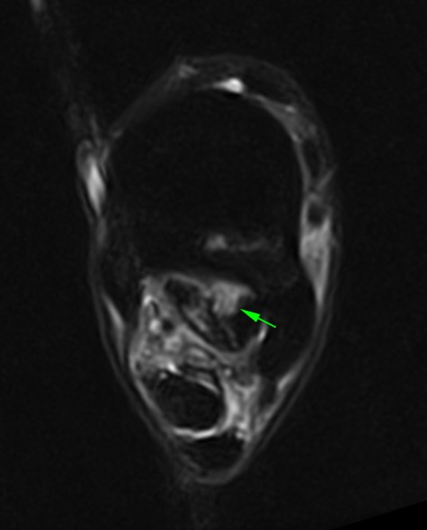
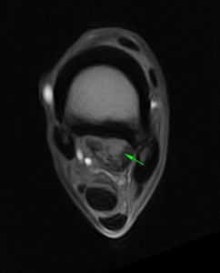
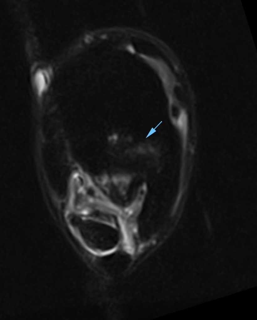
The horse underwent standing MRI of the left proximal metatarsal region. This revealed severe desmopathy of proximal aspect of the suspensory ligament with lesion of the dorsal fibrous part of the lateral lobe. There was bone edema-like lesion of the proximal plantaro-lateral aspect of the third metatarsal bone.
Conclusion
This case highlights the value of standing MRI in the assessment of proximal suspensory ligament injury, allowing detailed characterisation of both soft tissue and osseous pathology, including bone edema-like change within the proximal third metatarsal bone.
The use of iNAV motion correction enabled acquisition of high-quality images with excellent anatomical detail, even in the proximal metatarsal region, supporting accurate diagnosis and informed clinical decision-making.
With many thanks to Dr Matteo Toniato, Clinica Equina San Biagio, Italy for sharing this case with us.