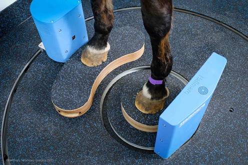


With more specialists under one roof than anywhere else in the country, the Dick Vet Equine Hospital is the most advanced equine hospital in Scotland. As the first university in Europe to install low-field magnetic resonance imaging (MRI) in 2005, Edinburgh were early adopters of imaging innovation. With over 130 Hallmarq Standing Equine MRI sites globally, Edinburgh are one of only seven to have achieved a 9.5 out of 10 for their image quality in 2025.
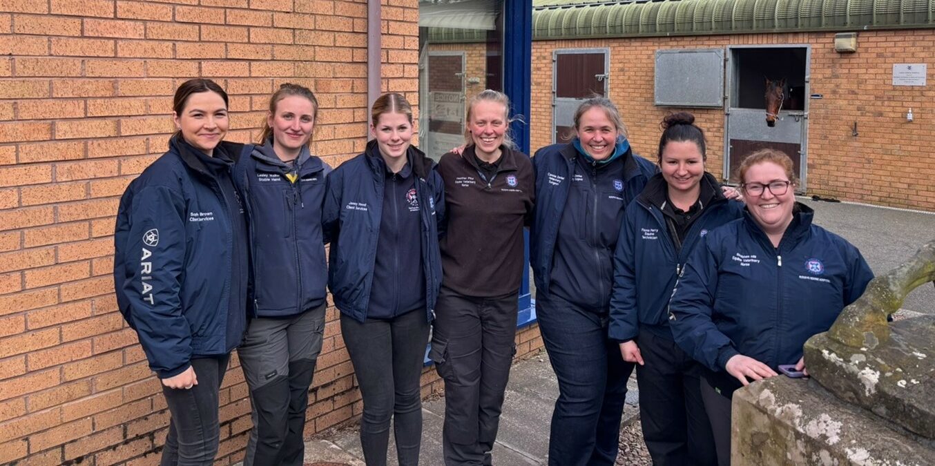
Dick Vets: Training Tomorrow’s Specialists
Over the past two decades, Standing Equine MRI has helped inform valuable research projects at the University, such as one of the cornerstone papers on the prognosis for deep digital flexor tendon (DDFT) injury within the foot (Cillan-Garcia et al 2013) [1]. A further project evaluated the relevance of fluid signal in the distal phalanx in horses with concurrent DDFT injury (Hewitt-Dedman et al 2021) [2]. Sequential projects evaluated the MRI appearance of naturally occurring cartilage injury of the distal interphalangeal joint on low-field MRI (van Zadelhoff et al 2020) [3], followed by a PhD project validating the use of T2 mapping on low field MRI with histology (Baker et al 2022) [4].
More recent projects now feed into the Vet School’s relatively new Professional Doctorate in Veterinary Medicine (Residency) program. Renowned and recognized for clinical and academic excellence, the initiative provides an opportunity for qualified veterinary surgeons to undertake a period of advanced clinical training in a chosen specialty under the guidance and supervision of the Royal College of Veterinary Surgeons, European and American boarded veterinary specialists.
The hospital’s purpose-built facilities also allow residents to access expansive multi-disciplinary training across radiography, CT, scintigraphy, ultrasound, and MRI. In close collaboration with Edinburgh’s senior clinicians, they not only gain hands-on expertise but contribute to world-class research. Indeed, many alumni now work in international specialist roles at referral centers and universities worldwide, testament to the Dick Vet’s impact beyond its own walls.
Why Combine Standing Equine MRI and Standing CT?
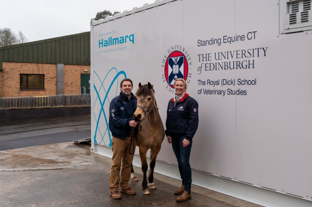
The decision to install Hallmarq’s Vision CT in 2024 was informed by a policy change at the University whereby the routine clench checks – formerly performed with radiographs – was replaced with Cone Beam CT (CBCT). Importantly, minimizing radiation exposure was a key consideration in the team’s decision making, with recent research by Gaida et al (2025) [5], on the lower cumulative radiation exposure of CT when compared to radiographs, providing further reason for the change.
Dr Padraig Kelly, Head of Dick Vet Equine Hospital, explains why the decision to undertake both MRI and CT scans for horses presenting with lameness, was made:
Offering both standing MRI and CT scans will significantly aid the diagnosis of lameness of our patients. Having both imaging modalities available provides an excellent opportunity for some sensitivity and specificity studies to determine whether CT or MRI is better for detecting different injuries in a horse’s foot.”
Dr Padraig Kelly, Senior Equine Surgeon, Director of the Dick Vet Equine Hospital, the University of Edinburgh
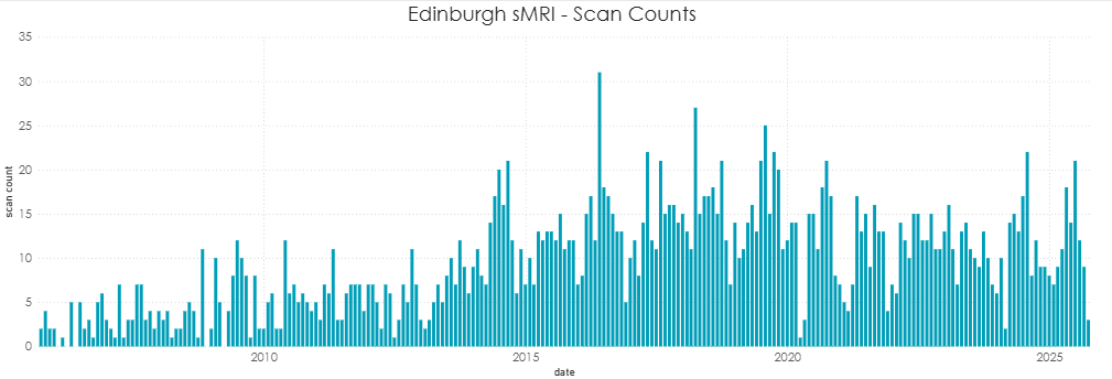
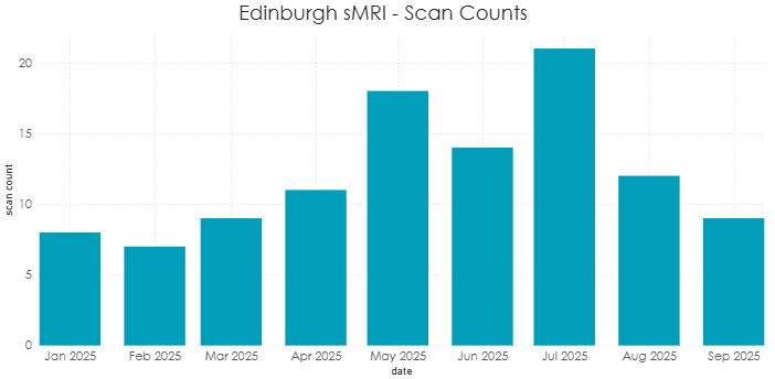
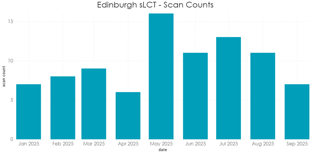
Exponential Growth
Scan volumes can and do vary across time particularly in the case of universities who are using their imaging modalities for valuable research. Caseload is far more than the next paying customer as data shows:
Evidence-Based Research
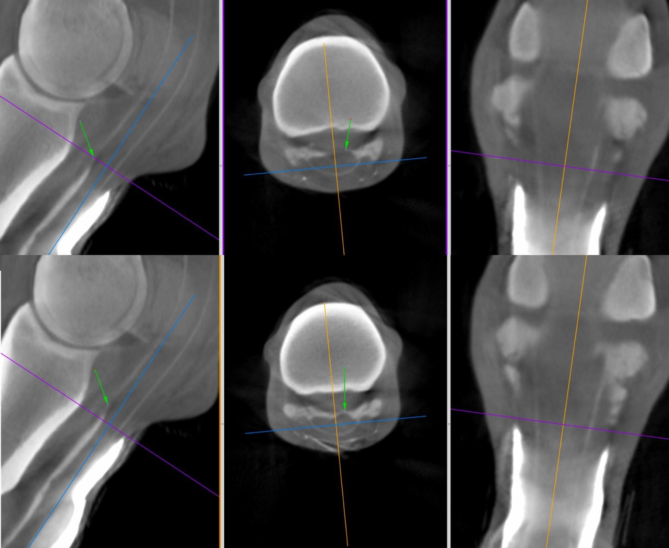
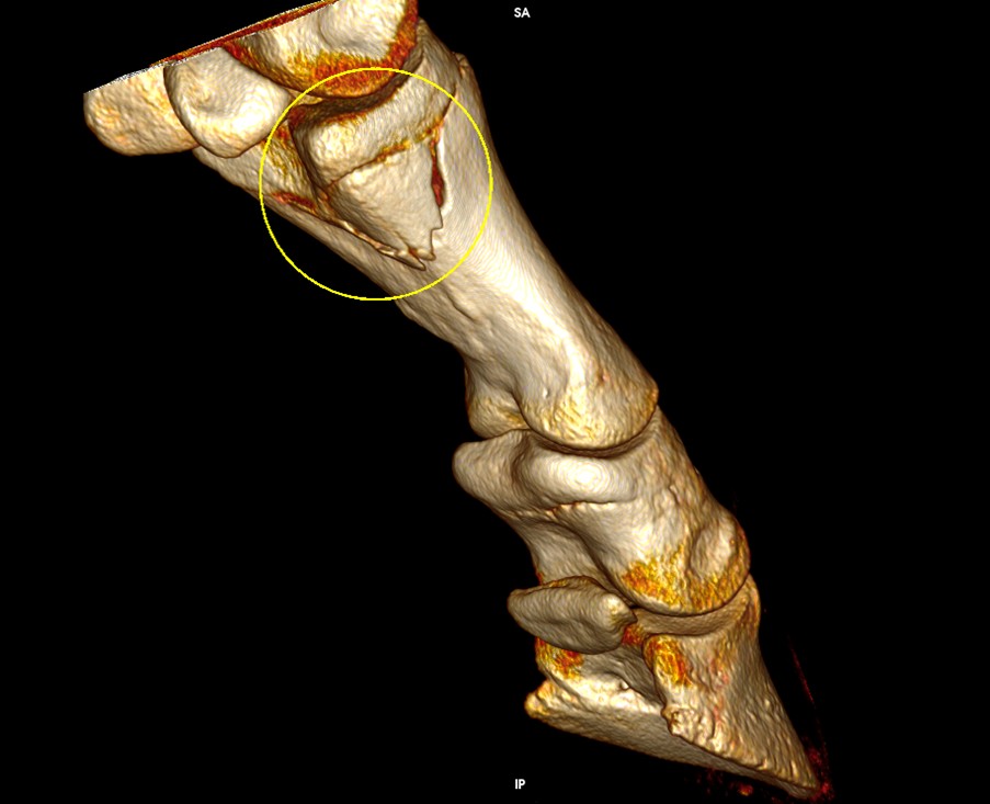
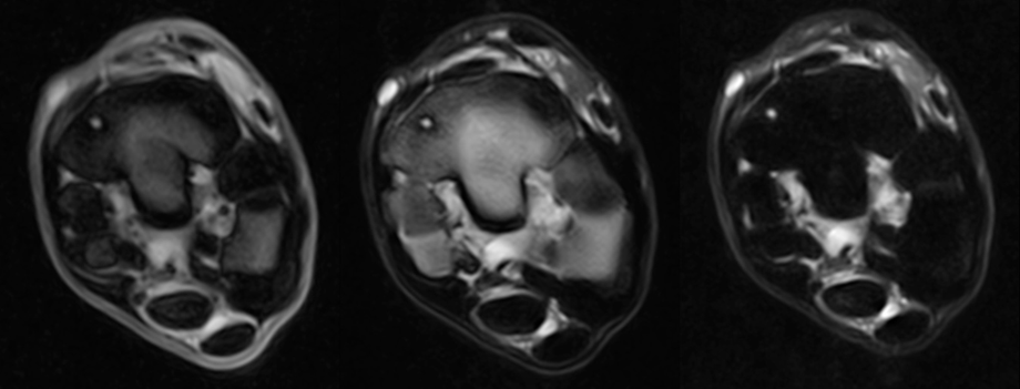
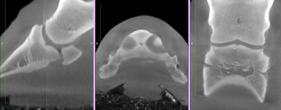
The Dick Vet’s pioneering research supports this dual-modality approach. In their 2025 study, Comparison of Cone-Beam CT and Low-Field MRI of the Distal Limb of 85 Standing Sedated Horses [6], the results were clear:
- 40% showed abnormalities on both modalities
- CT better detected subchondral bone lysis
- Maximum information is accrued when Cone Beam CT (CBCT) and MRI are used in combination
This case series identified findings evident on MRI that were not evident on Cone Beam CT (CBCT) in almost half of this case series. CBCT identified increased clarity of certain features such as the extent of subchondral bone lysis in this case of distal phalanx indentation. There were also instances where subchondral bone lysis was seen on CBCT but not on MRI albeit rarely. Taken together the maximum information is accrued when CBCT and MRI are used in combination increasing lesion detection, improving reader interpretation of images and providing increased assurance of correct diagnoses.
With the research ongoing for another 2 years, the use of a limited MRI protocol including STIR sequences for assessment of ‘active’ injury and CT scanning for structural imaging, there may be a possibility in the future to reduce scan times and optimize the acquisition of imaging information.
“Combining these advanced techniques has the potential to improve diagnostic sensitivity and specificity for difficult injuries within the equine foot.”
Dr Sarah Taylor (BVM&S MSc PhD Cert ES(Orth) DipECVS DipECVSMR FRCVS)
Senior Lecturer in Equine Orthopaedics, the Royal (Dick) School of Veterinary Studies, the University of Edinburgh

What Next for the Dick Vet?
Forging Ahead: The Role of CT in Evidence-Based Farriery
Aligned with the hospital’s wider three-year CT–MRI diagnostic research initiative, the University has recently secured funding for CT assessment of the hoof wall to assist farrier trimming. The program will lay the foundations for an evidence-based approach to hoof trimming with the long-term goal of supporting trimming protocols with quantifiable anatomical data. High-resolution, three-dimensional CT images offer detailed insight into hoof capsule architecture, specifically the deep and superficial layers in transverse orientation enabling farriers to identify asymmetries, wall thickness variations, and subtle structural anomalies that would previously have gone undetected.
The team is now building a dedicated CT image library focused on hoof wall conformation and how it relates to farrier intervention, between farriers and veterinary clinicians. Together, they are working to build a literature base that will complement the existing MRI-focused body of research.
Epitomising Equine Excellence
If success is measured in academic excellence, scientific integrity, and clinical progress, then the Dick Vet truly epitomize that criteria.
Their contribution, over many years, has undoubtedly led to the wider adoption of Standing Equine MRI with notable publications informing decision making. The recent move from radiography to CT, their evidence-based approach to dual-modality imaging, and their commitment to resident education makes them a true leader in equine diagnostics. For Hallmarq, this long-standing collaboration is more than a customer relationship, it’s a shared vision of improving horse health through better, safer, and smarter imaging.
To learn more about the Royal (Dick) School of Veterinary Studies at the University of Edinburgh, click here >>
References
- [1] Cillán-García, E., Milner, P.I., Talbot, A., Tucker, R., Hendey, F., Boswell, J., Reardon, R.J.M. and Taylor, S.E. (2013), Deep digital flexor tendon injury within the hoof capsule; does lesion type or location predict prognosis?. Veterinary Record, 173: 70-70. https://doi.org/10.1136/vr.101512
- [2] Hewitt-Dedman, C.L., Biggi, M., Van Zadelhoff, C., Schwarz, T., Reardon, R.J.M. and Taylor, S.E. (2022), Imaging findings and clinical outcome of foot pain attributable to insertional deep digital flexor tendon injury and/or fluid signal within the flexor surface of the distal phalanx. Equine Vet Educ, 34: e422-e430. https://doi.org/10.1111/eve.13503
- [3] van Zadelhoff C, Schwarz T, Smith S, Engerand A and Taylor S (2020) Identification of Naturally Occurring Cartilage Damage in the Equine Distal Interphalangeal Joint Using Low-Field Magnetic Resonance Imaging and Magnetic Resonance Arthrography. Front. Vet. Sci. 6:508. http://doi.org/10.3389/fvets.2019.00508
- [4] Baker ME, Kershaw LE, Carstens A, Daniel CR, Brown H, Roberts S, et al. T2 mapping of cartilage in the equine distal interphalangeal joint with corresponding histology using 0.27 T and 3.0 T magnetic resonance imaging. Equine Vet J. 2023; 55(5): 843–852. https://doi.org/10.1111/evj.13900
- [5] Gaida, J.L., Steinberg, T., Stieger-Vanegas, S.M., Merle, R. and Lischer, C.J. (2025), Equine Standing Multidetector Computed Tomography of the Distal Thoracic Limb and Tarsus Has a Lower Cumulative Radiation Dose than Digital Radiography. Vet Radiol Ultrasound, 66: e70049. https://doi.org/10.1111/vru.70049
- [6] Sarah Taylor, Padraig Kelly, Carola Daniel, et al. Comparison of cone beam CT and low-field MRI of the distal limb of 85 standing sedated horses. Authorea. May 19, 2025. https://doi.org/10.22541/au.174767603.34807364/v1