


Despite being one of the most prevalent spinal diseases among large and giant breeds, there’s still a lot of confusion over cervical spondylomyelopathy (CSM). The uncertainty isn’t helped by the disease being known by as many as fifteen different names!
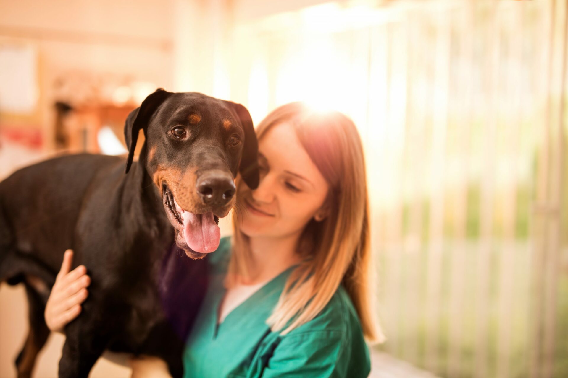
Most commonly affecting Dobermans and Great Danes, cervical spondylomyelopathy causes spinal cord and nerve compression. It generally manifests as either disc-associated or osseous-associated CSM and is known to cause neck pain (among other clinical signs) in giant and large breed dogs.
As part of our 2021 Summer Learning series, Hallmarq invited Dr. Ronaldo da Costa, a leading expert on disc-associated cervical spondylomyelopathy, to demystify this extremely common yet often misunderstood spinal disease. Here’s what you need to know.
How Can We Diagnose Cervical Spondylomyelopathy?
The number of affected dogs may be much larger than we realize. In 2006 Dr. Da Costa conducted a study of sixteen Dobermans. He found that 25% had spinal cord compression, 75% had disc degeneration and each of the subjects had some degree of disc protrusion, despite all the dogs presenting normally. Dr. da Costa performed the same study on Great Danes. Both studies are available for reference.
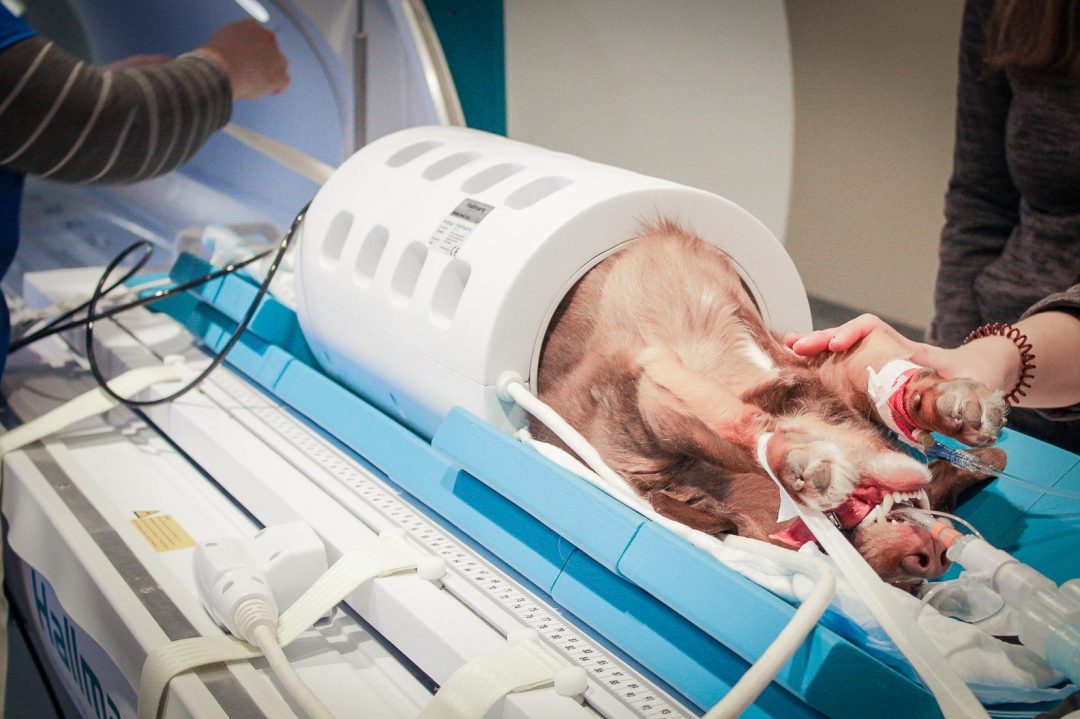
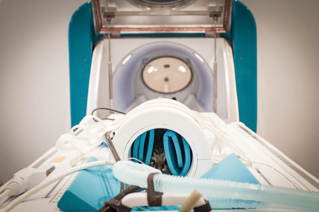
MRI is the best modality for diagnosing cervical spondylomyelopathy, but the condition is still easy to overlook. The disease is most prevalent in the caudal cervical spine, most often affecting C5-C6 and C6-C7. However, it is good practice to image the entire cervical spine. If you only focus on imaging C5 to C7, you can miss severe compressions in other parts of the spine. Always include C1 to T2 to increase the chances of a correct diagnosis. Compression between T1 and T2 is most common in giant breed dogs but can happen in large breeds as well.
Extension Helps You See the Severity of Compression
You need to put your patients’ necks in extension when imaging to observe the extent of cervical spondylomyelopathy. In dorsal recumbency (the standard position for spinal imaging), you won’t be able to witness the level of compression in the spine and neck. After all, dorsal recumbency isn’t a normal position for dogs, meaning the severity of compression won’t be as visible. Instead, you need to position your patients in ways they would normally hold themselves so you can see the full impact of the disease.
Additionally, cervical spondylomyelopathy often has a dynamic component. So it is useful to compare a neutral neck position with images of the neck when the patient’s neck is in extension; this allows you to see multiple dynamic compressions. Quite simply, compressions that are extremely apparent when the patient is put in extension can be easy to miss in a neutral position.
Avoiding Traction Myelography for Cervical Spondylomyelopathy Patients
Traction myelography can be harmful to the patient and may result in post-myelographic seizures and temporary deterioration of neurologic status. This promotes a reluctance to recommend it even if it was an informative option. This is reinforced by the fact that kinematic extension provides better information than traction, while also being less risky. Dr. da Costa included traction in his research and discovered its drawbacks. The studies he conducted show that using traction to image and diagnose cervical spondylomyelopathy can result in extremely inconsistent results.
If you do use traction in your advanced imaging studies, take care not to use more than 20% of the patient’s weight in traction. Anything above 25% becomes dangerous to the patient.
We still have much to learn regarding cervical spondylomyelopathy, but Dr. da Costa’s detail-rich research deepens our understanding.
To learn more, watch the complete webinar.