

Technology
How it works
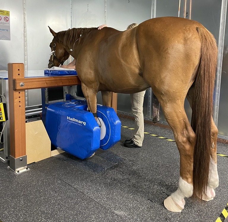
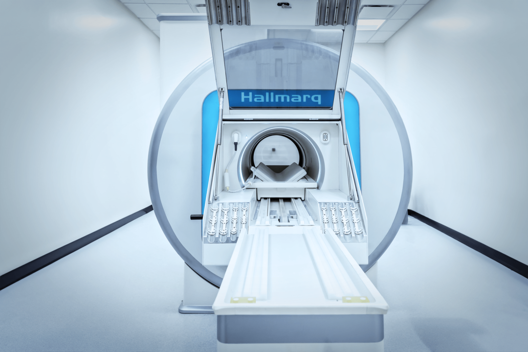
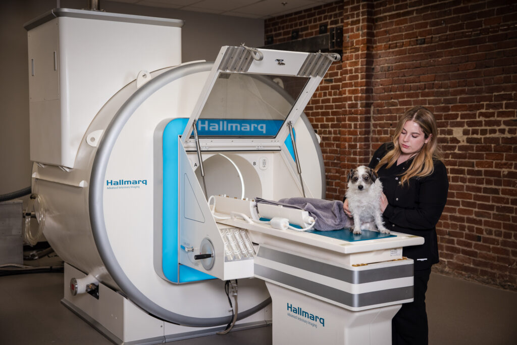
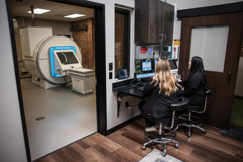
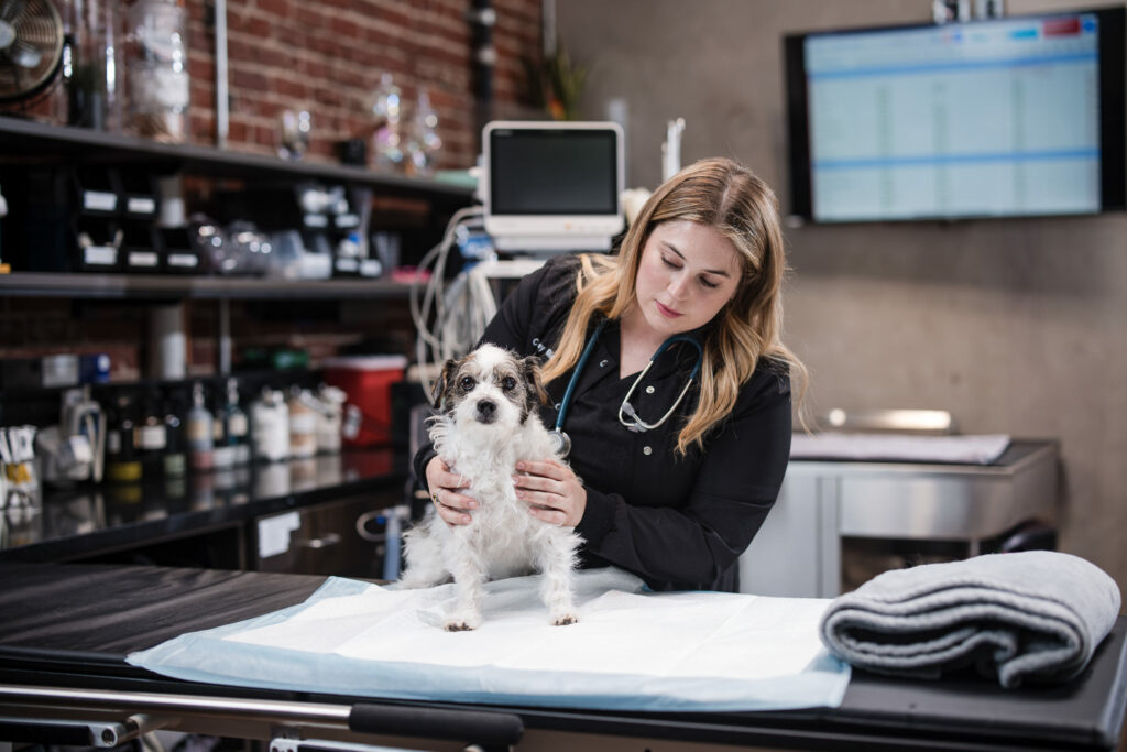
Hallmarq’s Small Animal 1.5T MRI is a cost-effective, flexible, and user-friendly imaging solution designed specifically for veterinary practice. Passionate about image quality, Hallmarq developed their machine to deliver superb detail for a definitive diagnosis. Its zero-helium technology, and built-in RF shield ensure that the cost and complexity of offering 1.5T MRI are minimized at installation and beyond.
The 16-channel self-shielded design offers an array of veterinary-optimized RF coils for proper patient positioning and enhanced image acquisition. With a V-shaped patient bed for increased scan coverage, and veterinary-optimized MRI software, Hallmarq’s 1.5T MRI delivers high-resolution detail to help diagnose and treat neurological conditions confidently and efficiently.
Details
Clinical uses
Spinal cord disease
Including spinal cord neoplasia, intervertebral disc disease, wobbler’s disease and lumbosacral disease, atlanto-axial subluxation, ischemic myelopathy, nerve root compression and discospondylitis.
Brain disease
Including, but not limited to neoplasia, cerebrovascular events, hydrocephalus and inflammatory brain disease.
Skull and surrounding soft tissues
The only modality able to completely assess otitis externa-media-interna, including assessment of associated cranial nerves, cochlea and semi-circular canals along with the adjacent brainstem. Nasal and orbital disease can also be clearly assessed.
Cranial and vertebral anomalies and genetic malformation screening
Assessment of congenital anomalies including vertebral malformations, Chiari-like malformation, and syringomyelia screening in pre-disposed breeds.
Stifle disease
Superior soft tissue imaging for assessment of cranial cruciate ligaments, meniscal injuries and muscular disease.
Shoulder disease
Superior soft tissue imaging for assessment of biceps and supraspinatus tendon pathology, joint capsule, brachial plexus disease and muscular disease.
Thoracic and abdominal disease
Complimentry to ultrasound and computed tomography, soft tissue diseases affecting the lungs, liver, kidneys, spleen and major vessels can be accurately assessed with MRI.
Epilepsy screening
Confirmation of idiopathic epilepsy requires an imaging investigation to rule out structural causes, toxicities and metabolic diseases. If an MRI of the brain is considered normal, when accompanied by a normal CSF analysis, the cause of seizures is compatible with an idiopathic disease.

Why MRI?
Features & benefits
NO helium, NO quench pipe, NO oxygen monitor required
Our unique zero-helium design delivers reduced upfront and ongoing costs with a less complex installation.
Veterinary software and protocols
Included. You don’t have to rely on outsourcing to develop protocols or use sequences designed for humans.
Veterinary-specific coils
With an over-sized v-shaped spine coil (91 cm) and elongated head coils with removable tops, signal is increased by 20% while allowing better patient access.
Affordable
Capped monthly payments help standardise and stabilise your overall cost of ownership and reduce the operational risks of implementing an MRI service.
99% uptime guarantee
Decrease costs, avoid wasted time and increase customer satisfaction with our 99% uptime guarantee as standard.
Unique self-shielded hatch
Our built-in RF shield reduces the cost and complexity of installation while allowing free access to your MRI room with video monitoring to increase patient safety.
Dual coil functionality
Our dual coil system provides a significant boost in signal intensity across larger body regions allowing fast scan times and pristine images of exceptional diagnostic quality.
Q-Care Support
From initial enquiry through system installation and beyond, you’ll receive unparalleled support from a team dedicated to the veterinary profession.
Flexibility
Installation
If available space prohibits an in-hospital MRI suite, our modular and trailer options offer the perfect solution. Delivered and set up in days, our MRI trailer is a reliable, modern way to expand your advanced imaging service. For a more permanent option, our custom modular building provides a structure with ample room. Either way, there’s no compromise. Both deliver the same superb image quality you’d expect from an in-hospital MRI suite
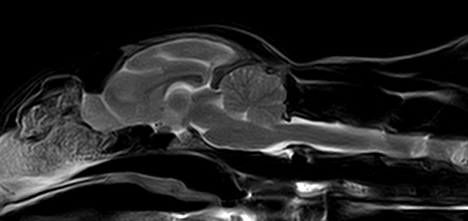
Detailed imaging
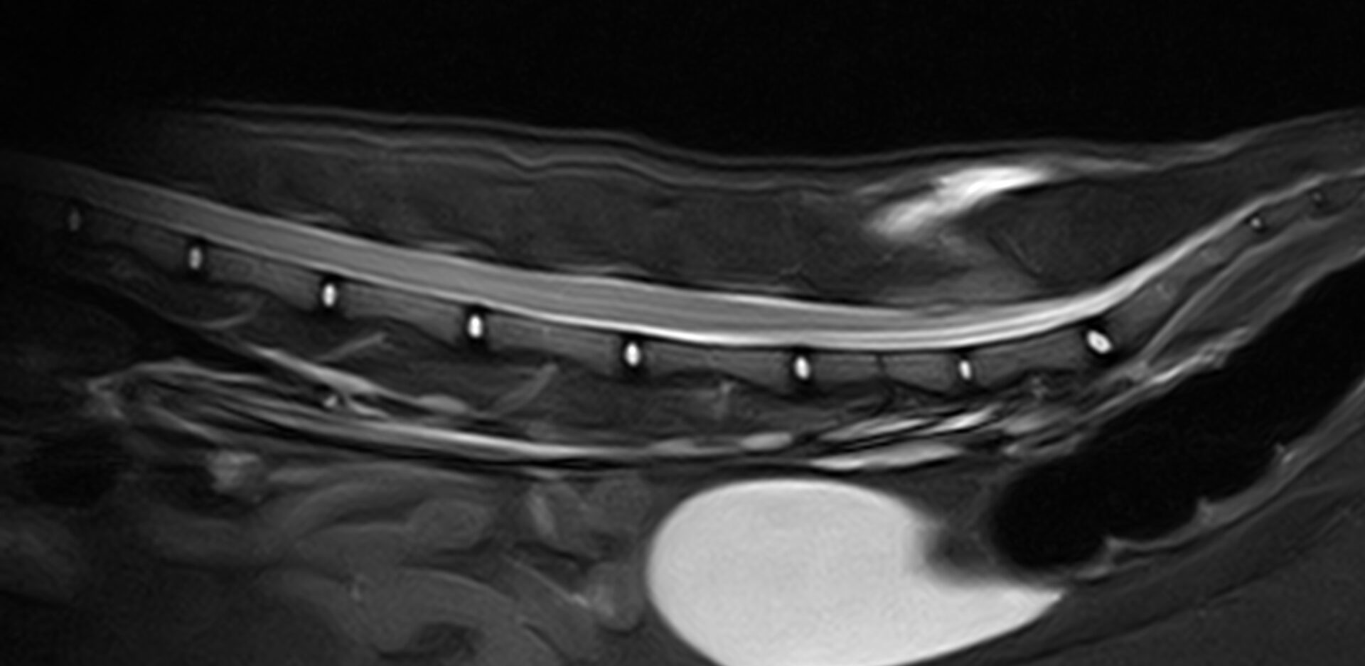
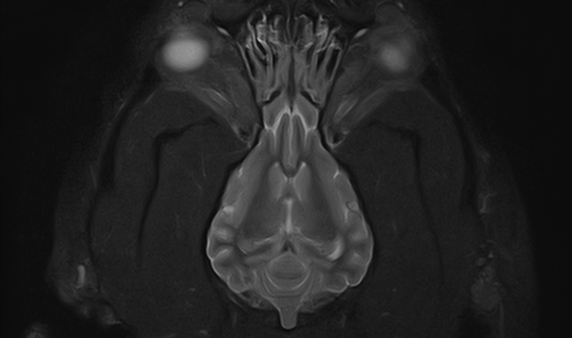
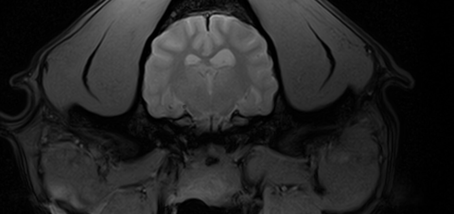
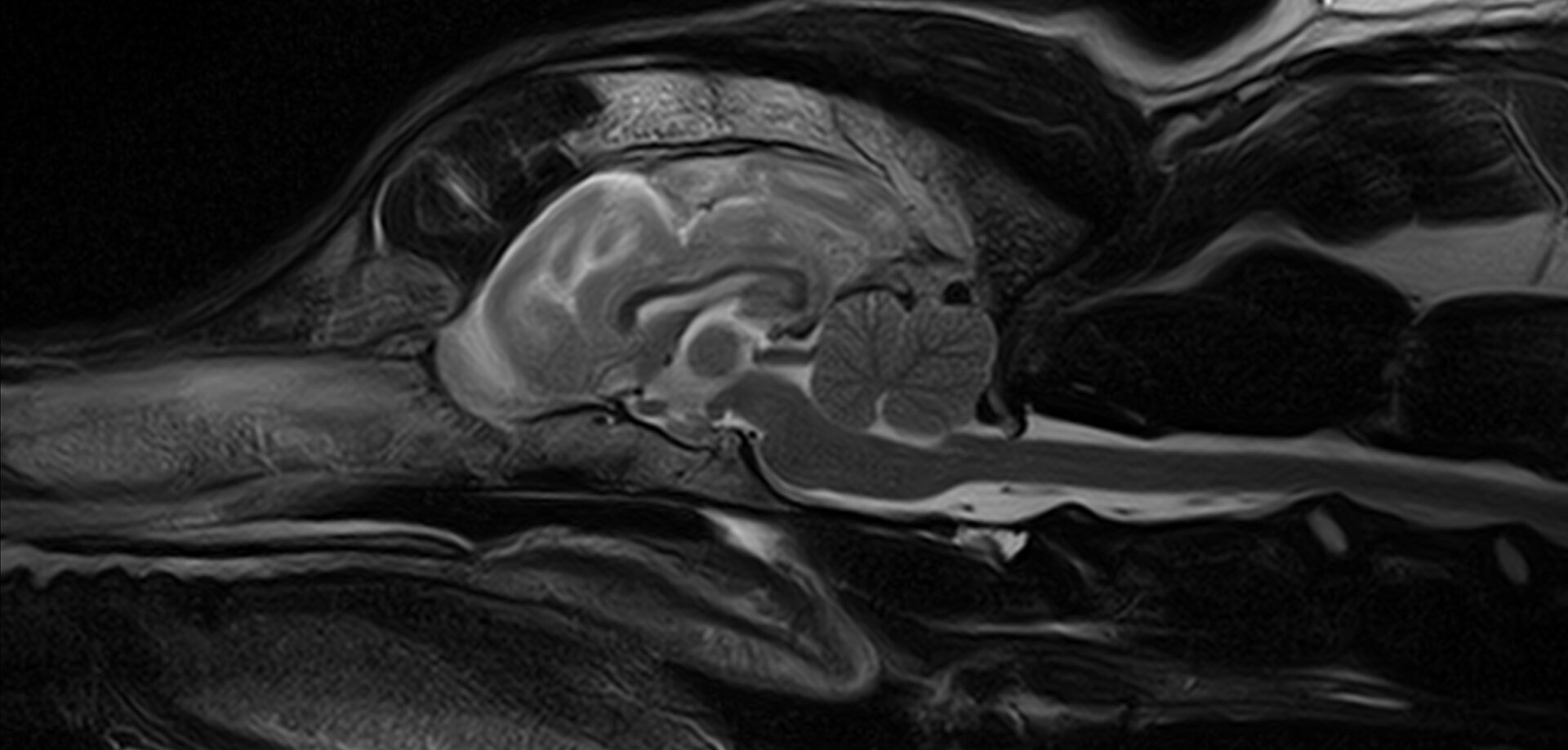
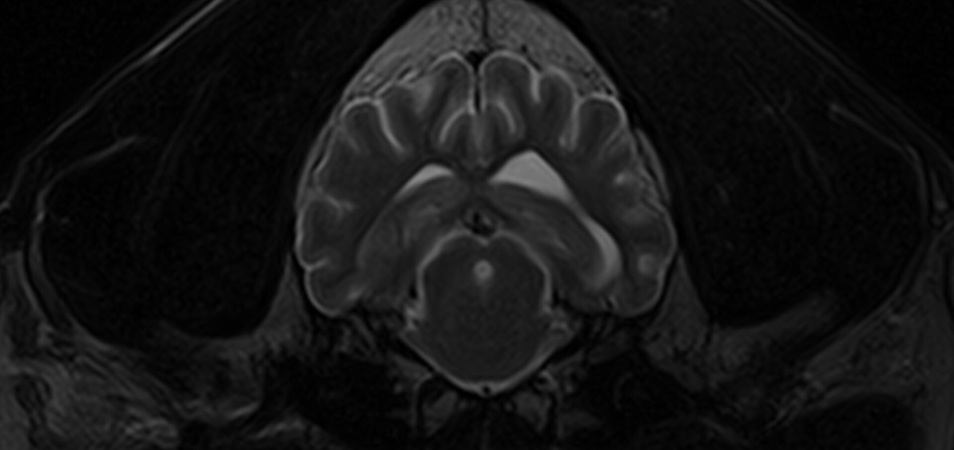
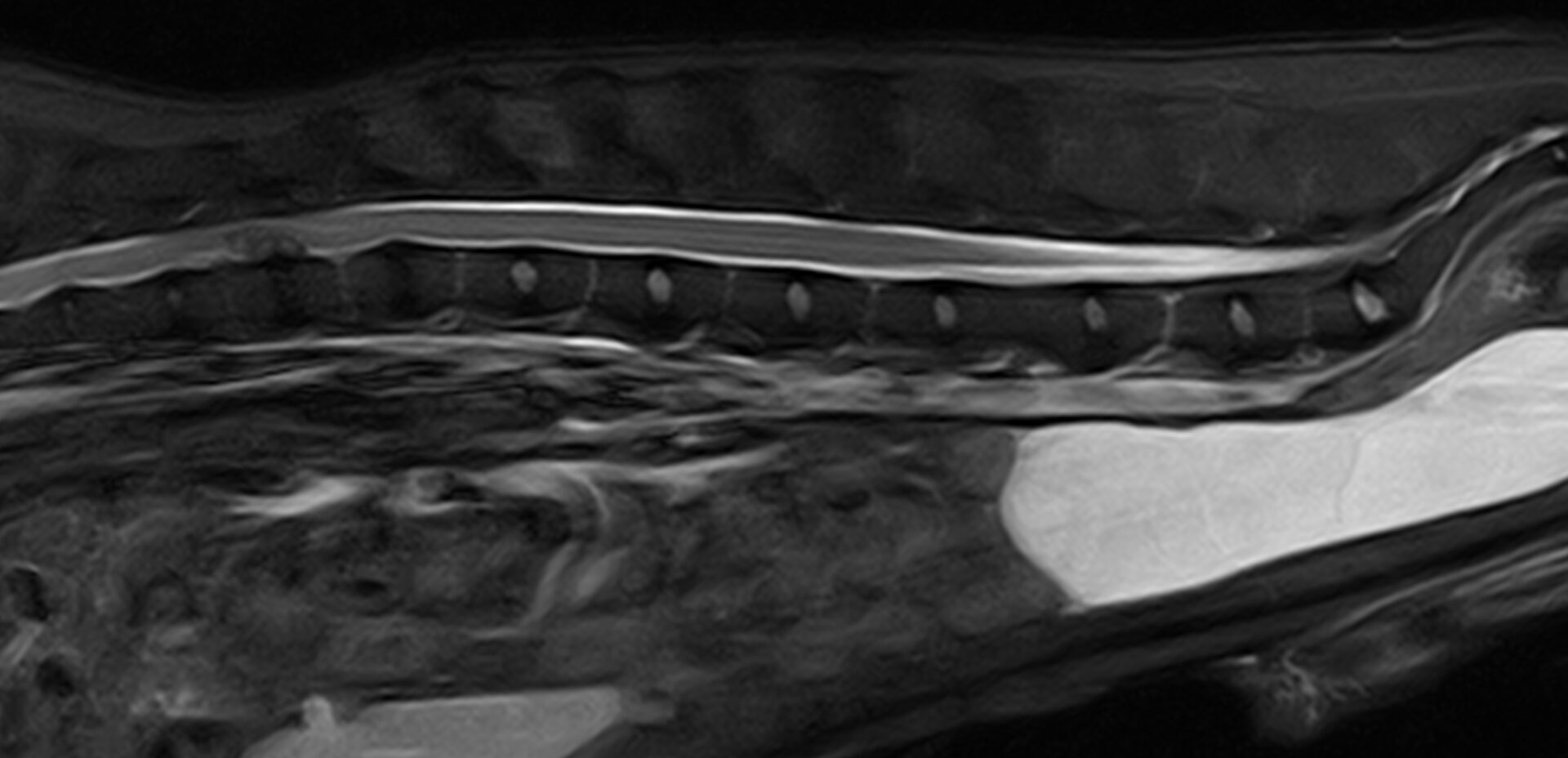
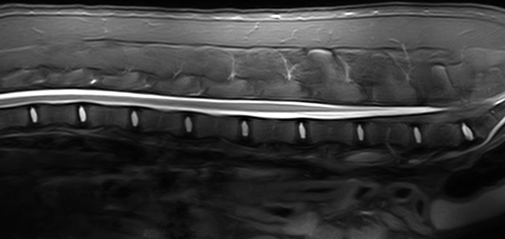
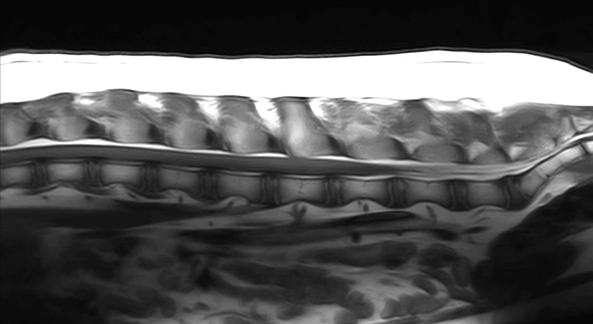
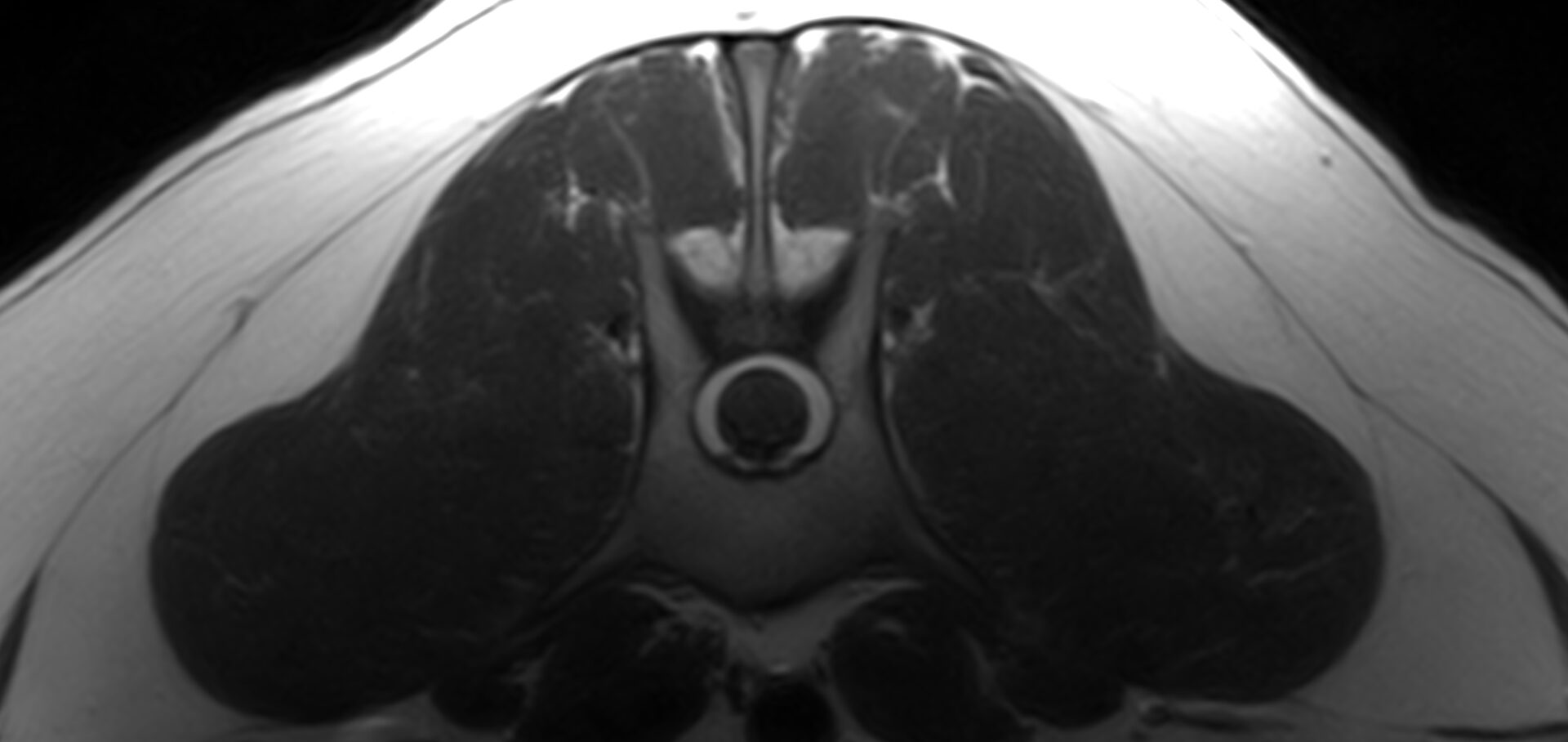
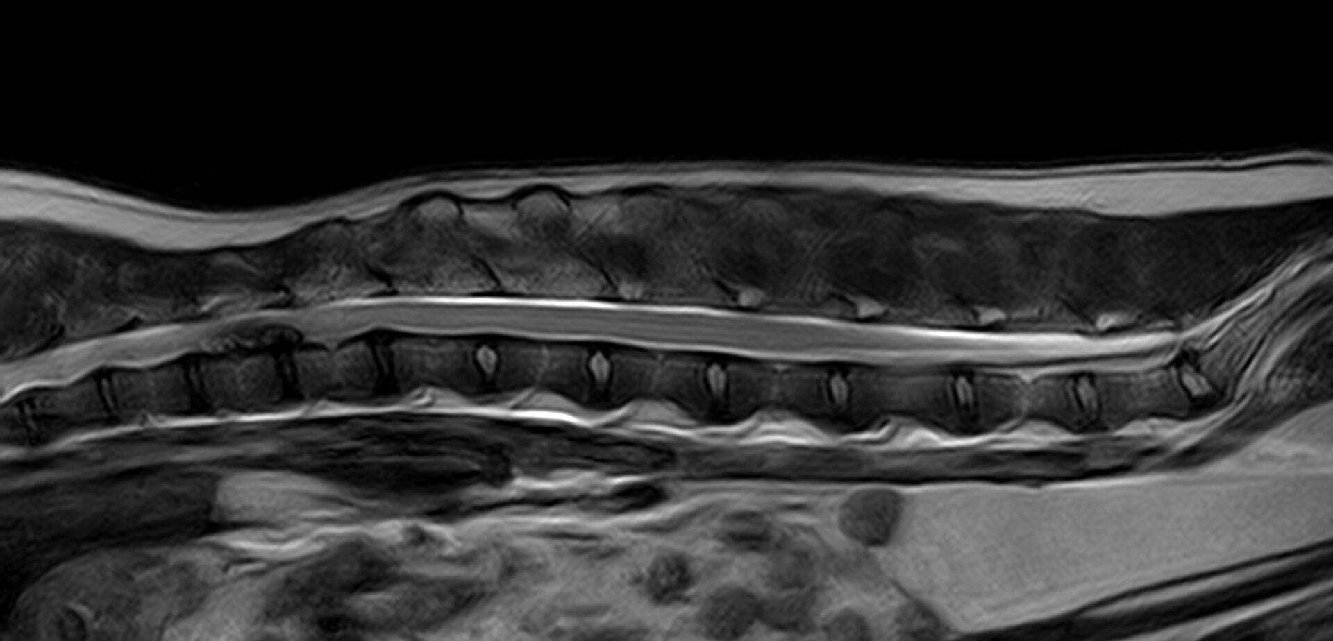
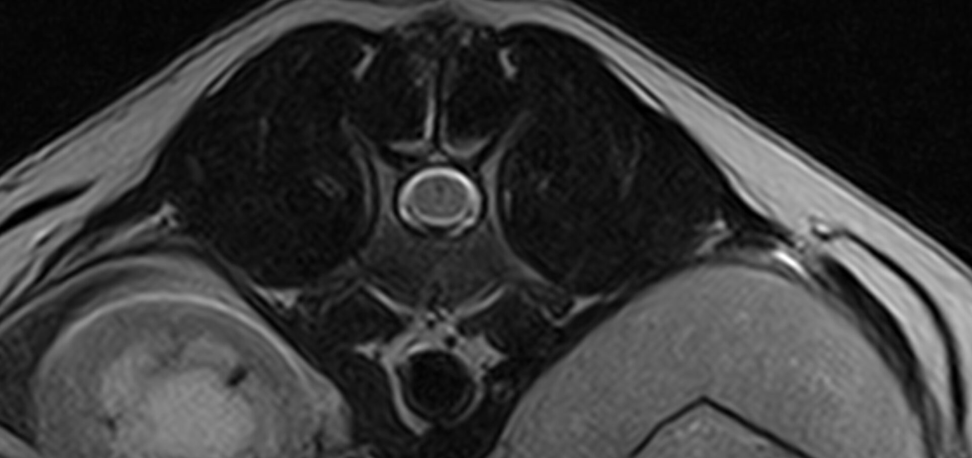
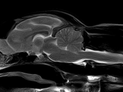
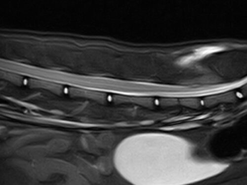
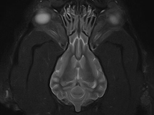
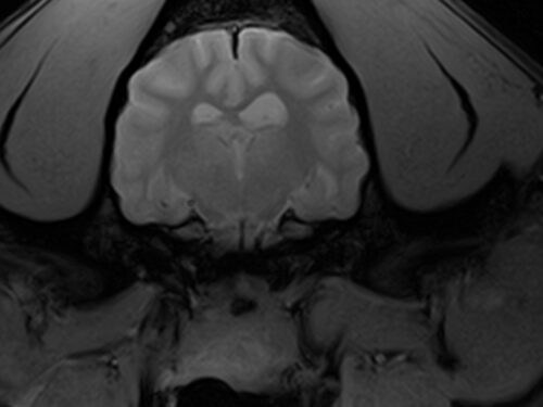
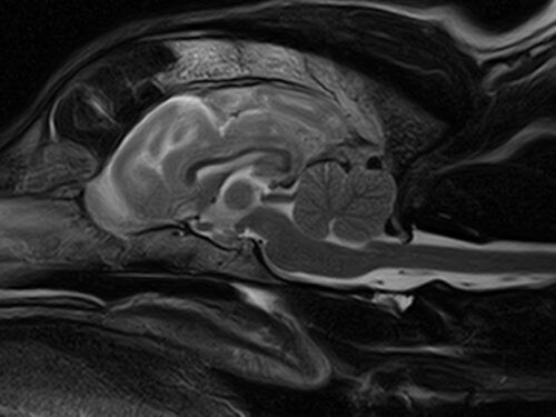
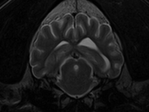
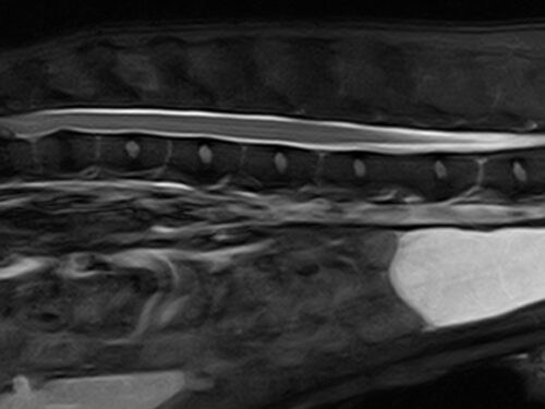
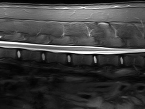
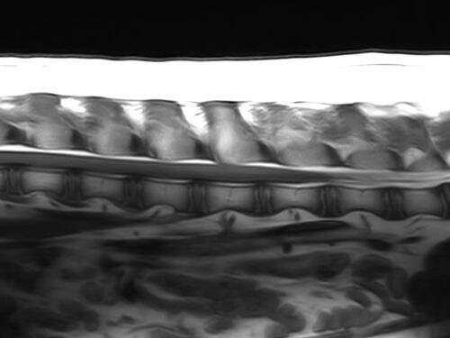
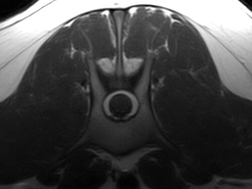
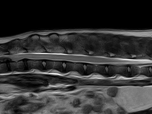
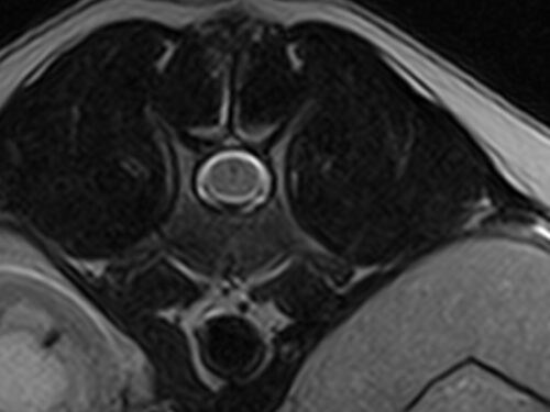
Clinical gallery
Specifically designed to image small animal anatomy, Hallmarq’s zero-helium 1.5T MRI captures superb images for a more accurate diagnosis.
- 16-channel, self-shielded design: an array of veterinary-optimized RF coils help to properly position the patient
- Enhanced RF coil coverage: for superior image quality when compared to equivalent human-optimized RF coils
- Extended V-shaped phased array spine coil: used in combination with our 8-channel flexible coil, increases scan coverage for larger breeds, enhancing image quality for difficult-to-scan regions, such as the brachial plexus
- Veterinary optimized MRI software: superb high-resolution detail including pre-saturation, fat suppression, blood flow compensation, single-shot fast spin echo, intensity correction and diffusion imaging with automatic ADC maps
Hallmarq’s ability to use different coils and positioning to capture images is probably unparalleled.”
Dr. Billy Pullen, DVM, DACVIM (SAIM) Veterinary Care & Speciality Group, Chattanooga, TN
I expect to see our caseload grow tremendously as word gets out about our neurology service and MRI capabilities.”
Dr. Baye Williamson, DVM, DACVIM Veterinary Emergency and Referral Center (VERC), Oahu, Hawaii
Hallmarq’s 1.5T high-field magnet with its own shielding is revolutionary. It provides us with a lot more flexibility to look at our patients in greater detail.”
Dr. Christiane Massicotte, DVM, MS, PhD, DACVIM (Neurology) Veterinary Care & Speciality Group, Chattanooga, TN
I worked with a 1.5T Siemens and GE machines prior to the Hallmarq unit. Comparing these machines of the same magnet size, Hallmarq’s machine creates images that are by far superior in quality and clarity which I have always attributed, to some degree, to the veterinary specific coils.”
Dr. Dana Gietzen, DVM Las Vegas Veterinary Speciality Center
The addition of the Hallmarq MRI significantly improves our ability to diagnose and treat pets with brain and spinal cord abnormalities.”
Dr. Erika Sox, DVM, DACVIM Veterinary Emergency and Referral Center (VERC), Oahu, Hawaii
I regularly review brain MRIs from other referral hospitals for second opinions. I’m very impressed with the quality of our Hallmarq MRI images relative to traditional used high-field MRIs. The clarity of our images strengthens our confidence in central nervous system disease diagnosis.”
Dr. Cody Alcott, DACVIM Tucson Veterinary Specialists, Tucson, AZ
Hallmarq’s 1.5T MRI is designed specifically for small animal anatomy and delivers accurate and detailed images with great image resolution and contrast. The essential safety features and operator friendly software make it an asset to any veterinary neurologist.”
Dr. Franziska Fitz, DVM, GPCertSAS, DACVIM (Neurology/Neurosurgery) Las Vegas Veterinary Speciality Center
At VCSG, we are very pleased with the image quality our Hallmarq MRI equipment provides. It has allowed us to give more specific diagnoses and state of the art treatment recommendations for our patients. The technical support from Hallmarq is also excellent.”
Dr. Massicotte, DVM, MS, PHD, DACVIM (Neurology) Veterinary Care & Speciality Group, Chattanooga, TN

Cost
Significant upfront investment is a major concern for most veterinary hospitals. With the Hallmarq solution, you don’t have to worry about securing a bank loan to purchase the equipment; a single monthly payment covers everything. Furthermore, with no costly helium refills, no RF shield and MRI Techs available to remotely operate your system if needed, your savings are considerable.
Complexity
We appreciate that installing 1.5T MRI can be a complex process. That’s why the challenges of installing, running, servicing, and supporting an MRI machine have been carefully addressed by our team. The machine’s built-in RF shield eliminates the need for an expensive room build out and decreases building works. And, because Hallmarq’s 1.5T MRI contains no helium, there are no extra costs and permit issues associated with installing a helium quench pipe.
Commitment
Deciding to go ahead with 1.5T MRI is a big commitment. Which is why we’ve put together the total advanced imaging package to support your business decisions.
“Hallmarq is a one stop shop. I didn’t need to worry about finding a buyer of a used magnet on the secondary market, find an installer, and find someone to maintain the machine; Hallmarq produces, delivers, installs, and maintains the equipment and software used to run the equipment.”
Dr Adam Moeser, DVM, DACVIM (Neurology) Wisconsin Vet Neurology and Surgical Center
FAQs
Frequently asked questions
- Is high-field MRI expensive?
- Can I speak to an end-user?
- How much space should I allocate?
- How good is Hallmarq’s zero-helium Small Animal 1.5T MRI image quality?
- What is the uptime percent of the Hallmarq zero-helium Small Animal 1.5T system?
- Is this simply a refurbished human magnet?
- We’ve decided on your zero-helium Small Animal 1.5T MRI system. How long do we wait before scanning our first patient?
The total cost of ownership is less than you may expect. With Hallmarq’s zero-helium Small Animal 1.5T MRI, you can either buy the system outright or rent using our unique Flat Rate model. With our built-in RF shield, there is no need for a purpose-built shielded room; upfront capital expenditure is kept to a minimum and there is NO helium required. We’ve designed the system, from the ground up, to save on energy and our comprehensive Q-Care support programme even includes software updates. Labour costs may also be minimised as the system is designed to be operated by a veterinary technician or nurse rather than a dedicated MRI Technologist.
Find a System
Our systems are available at some of the world’s leading veterinary facilities. To find a Hallmarq MRI or CT machine near you, click below.
Find a siteDetails
Specification
This outlines some of the high-level technical specifications and considerations in planning for the installation of the Hallmarq Small Animal 1.5T MRI machine. Please consult the Site Planning Guide for additional details.
| Key features | |
|---|---|
| Field System | State-of-the-art veterinary specific 1.5T high field system |
| Spine coil | V-shaped spine coil for 20-percent better signal to noise ratio |
| Image protocols | Veterinary image protocols designed by MRI experts |
| Hardware | |
|---|---|
| Field strength | 1.5T |
| Channels | 16 |
| Energy efficiency | Up to 25% more energy efficient compared to used MRIs |
| Bore diameter | 60 cm patient bore diameter |
Q-Care
Supporting Your Success
Providing high-quality images is just the beginning. With an unrivalled program designed to add value to your practice, we’re here to help optimize your imaging service – from initial enquiry through installation and beyond.