


Introduction
The field of equine health has always embraced technological advancements in diagnostic imaging that play a pivotal role in elevating the standard of care for our equine companions. From the original wet chemistry radiographs to the sophisticated technologies of today, each progression has further illuminated our understanding of equine anatomy and pathology. And, in this dynamic and fast-moving arena, Hallmarq Veterinary Imaging has emerged as a leading innovator in imaging technology, heralding a new era in veterinary diagnostics with each development.
Staying Ahead of the Curve
From the outset, Hallmarq has been at the forefront of advancements in equine diagnostic imaging. The company’s pioneering work in developing the world’s first, and only, Standing Equine MRI machine paved the way for more practical, clinic and horse-friendly solutions.
The Vision CT scanner is testament to Hallmarq’s ongoing mission to make advanced imaging not only more accessible but also practical and safe. With its distinctive design and application, the information that Vision CT provides helps address the clinical need in veterinary medicine for accurate lameness diagnosis with minimal risk to the horse. Its technology was developed with a clear understanding of the everyday challenges faced in vet practices and the paramount importance of safe, effective, affordable imaging.
As we explore the advantages and capabilities of Vision CT, we will see that the technology is so much more than mere advancement in diagnostic imaging – it represents a significant leap in offering more compassionate equine care. Hallmarq’s commitment to veterinary excellence is seen in every facet of Vision CT making the scanner a desirable asset for every equine practice.
The Current State of Equine Diagnostic Imaging
Equine diagnostic imaging has undergone considerable transformation, shaped by a combination of technological advancements and a deepening understanding of equine health. This journey, marked by significant milestones, has seen veterinary professionals adapt and embrace a variety of imaging modalities, each bringing its unique value to the diagnosis and treatment of equine conditions.
Veterinarians rely predominantly on radiography and ultrasound as first-line diagnostic imaging tools. These methods, while invaluable, have their limitations. Radiographs, for instance, offer a clear view of most bony pathology but fall short in detailing soft tissue injury. Conversely, ultrasound excels in soft tissue imaging but cannot penetrate further than the bone surface. This dichotomy often requires the use of both modalities concurrently to form a comprehensive diagnosis, leading to increased time, cost, and complexity for the veterinarian, horse, and owner.
Hallmarq’s Vision CT – Redefining Equine Diagnostic Imaging
The arrival of Hallmarq’s Vision CT is helping set a new standard within the field of equine diagnostic imagining. Its innovation stems from a profound understanding of the inherent challenges and limitations faced with traditional imaging methods, coupled with a forward-thinking approach to veterinary care. Vision CT represents not just an evolution in technology but also a paradigm shift in how equine patients are diagnosed and treated.
A cornerstone of Vision CT’s design philosophy is its capability to perform scans on the standing sedated horse. This feature moves away from the requirement for general anesthesia – a process that can pose a risk to the horse¹ and complicates the logistics of equine care. The ability to perform scans in a standing position not only mitigates these risks but also presents a more complete view of the horse’s anatomy under normal load-bearing conditions.
Vision CT – Streamlining Scanning
Vision CT enables fast capture of 3D image sets in just minutes and, with Hallmarq’s proven motion correction software designed to minimize artifact, you get so much more than a conventional radiograph. This is particularly advantageous when diagnosing conditions that are influenced by the horse’s posture or weight-bearing status, offering insights that were previously challenging to obtain.
Vision CT also excels in terms of operational efficiency. Traditional CT scanning can be a time-consuming endeavor requiring extensive preparation and recovery time, primarily due to the anesthetic needed. However, Vision CT streamlines this entire process. Its rapid scans reduce stress for the horse and cuts down acquisition time, allowing veterinary staff to focus on treatment rather than managing the complexities of imaging procedures. Crucially, this increase in efficiency does not come at the expense of image quality – Vision CT is engineered to produce high-resolution images with clarity and precision, offering superb insights into the equine distal limb.
The design of Vision CT prioritizes the safety and comfort of the horse, reflecting Hallmarq’s commitment to animal health. It ensures a stress-free experience for both horse and handler. This consideration extends to the user experience for veterinary professionals. Vision CT boasts a user-friendly interface and is designed for seamless integration into veterinary practices, irrespective of their size or specialty. This ease of use ensures that advanced diagnostic capabilities are accessible to a broader range of veterinary practices without requiring significant changes to their existing workflows or extensive training for their staff.
“We find Vision CT quick and easy to use. The horses tolerate the procedure quite happily and it provides a cost-effective option for the client.
Dr. Ellen Singer BA, DVM, DVSc., Diplomate ACVS & ECVS, FRCVS, American, European and RCVS Specialist in Equine Surgery, Veterinary Associate, Sussex Equine Hospital, UK.
In essence, Vision CT by Hallmarq is a thoughtful response to the nuanced needs of veterinarians and their equine patients. It stands as a testament to Hallmarq’s dedication to innovation, practicality, and the advancement of veterinary medicine, helping to set new standards in care and redefine what is possible in the diagnosis and treatment of equine lameness.
Benefits of Vision CT for Every Equine Practice
The introduction of Hallmarq’s Vision CT into veterinary practices offers a suite of benefits that extend beyond the immediate gains in diagnostic accuracy. This advanced imaging solution is helping redefine standards of care, enabling practitioners to offer services that were previously unimaginable in their precision and efficiency. The multifaceted advantages of Vision CT underscore its value as a pivotal addition to those veterinary practices committed to excellence in equine care.
Enhanced Diagnostic Accuracy:
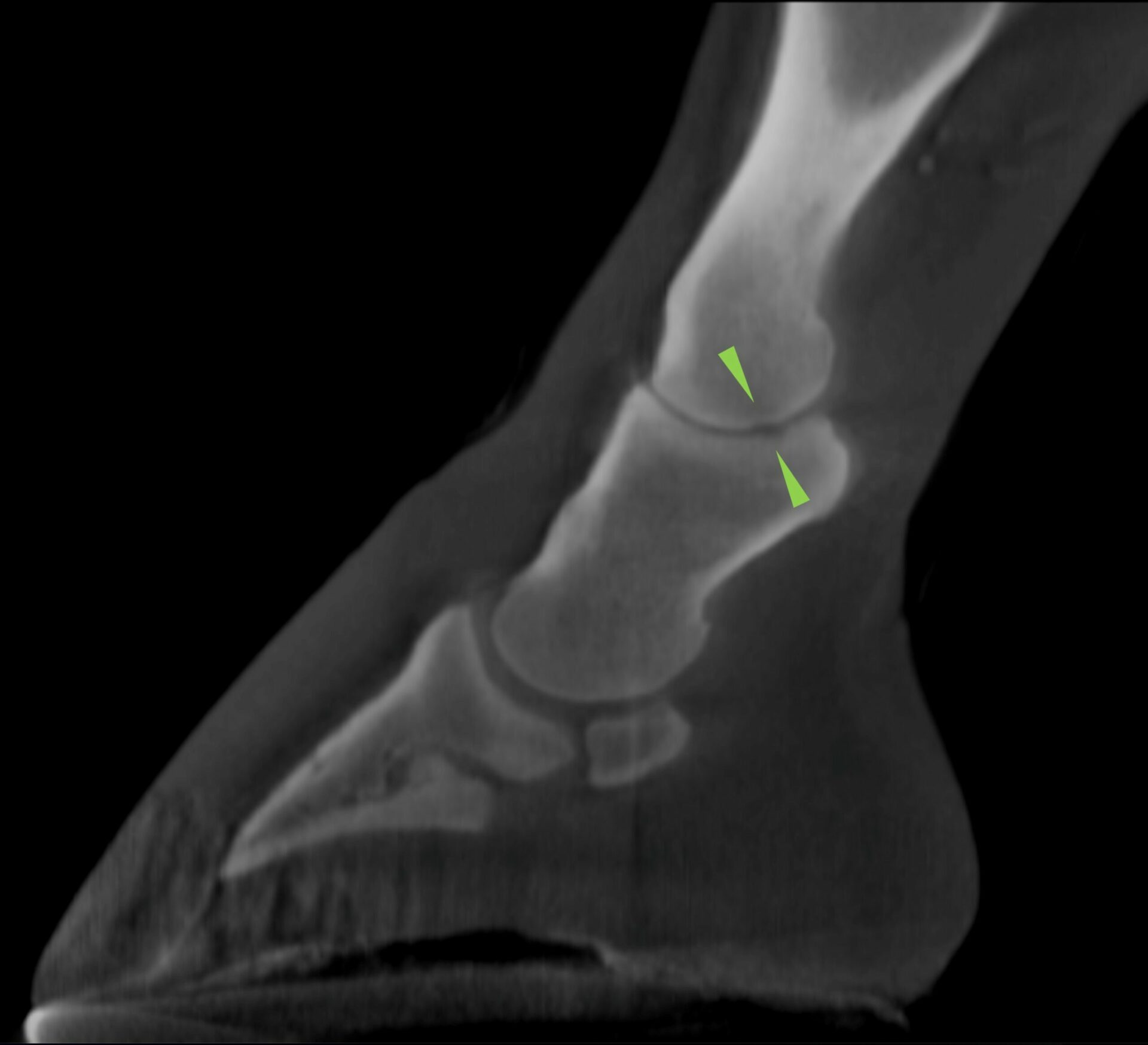
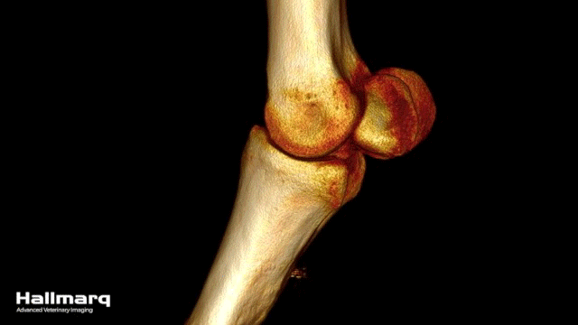
Vision CT offers superb spatial resolution to locate small changes in the bones of the distal limb, with no superimposition or overlap of anatomy.
“Standing Equine Leg CT is now our first port of call for complex proximal sesamoid fractures where we have concerns over configuration. Generating data sets is quick and easy and image resolution can be just as impressive as MRI.”
Henry O’Neill, MVB DVM MS Dipl ACVS MRCVS American Specialist in Equine Surgery, Donnington Grove Equine Vets, UK.
This accuracy is instrumental in diagnosing complex cases, where the limitations of radiography and ultrasonography might otherwise lead to inconclusive results. The technology’s capacity to capture high-resolution images in just minutes, coupled with the ability to orientate at any angle in standard workstation software to view key structures, translates into a deeper understanding of equine anatomy and pathology, empowering veterinarians with the information they need to make diagnostic decisions.
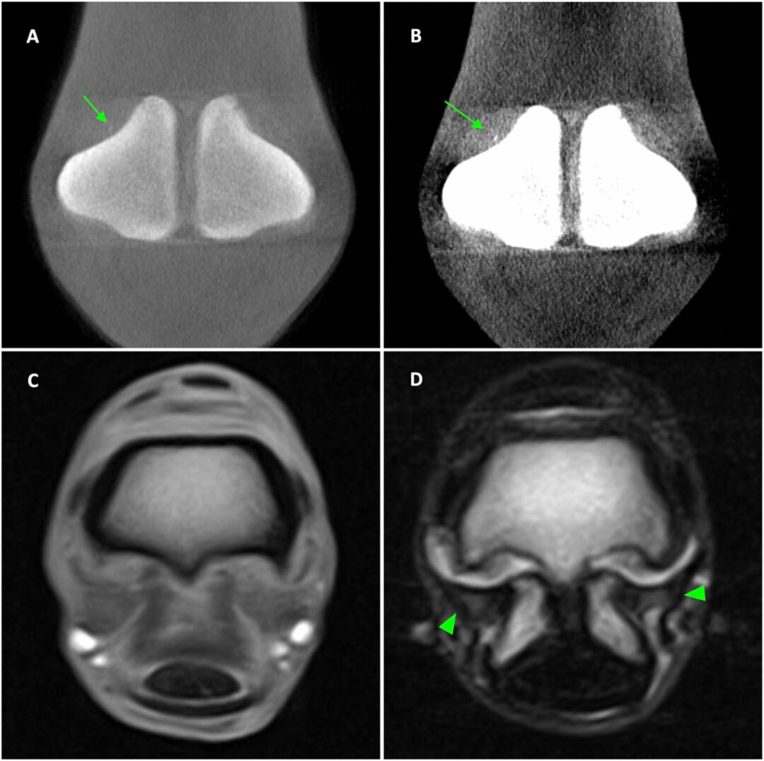
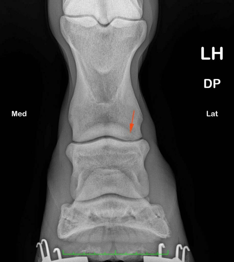
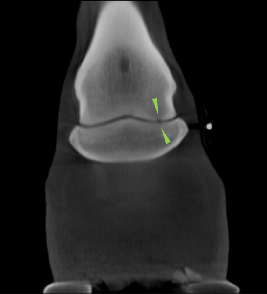
Figures 1 and 2 (below) show the same limb imaged with traditional X-ray (Fig. 1) and again with Vision CT (Fig. 2). While the radiograph image shows lucency in the lateral condyle of the proximal phalanx (orange arrows), imaging with Vision CT enabled a more quantitative assessment. In this case, a small and poorly marginated region of hypoattenuation of the adjacent trabecular bone (green arrows) can be seen. A shallower kissing lesion extends in the same direction through the subchondral bone plate of the lateral glenoid of the middle phalanx with no extension to the adjacent trabecular bone.
Increased Safety and Comfort:
One of Vision CT’s most significant features is the enhanced safety and comfort it offers to equine patients. Its walk-in-scan-walk-out standing capability eliminates the need for general anesthesia or awkward positioning, which reduces the associated risks and stress for horses, particularly those with pre-existing health conditions. This approach not only prioritizes animal health but also aligns with the growing emphasis on minimizing invasive procedures in veterinary practice.
Operational Efficiency:
Vision CT stands out with its operational efficiency, a feature that resonates strongly with busy veterinary practices. Its rapid scanning capability significantly reduces the time required for diagnostic procedures, allowing practices to accommodate more patients and improve the overall quality of care. This efficiency does not compromise the quality of diagnostic information though. Instead, it enhances the practices’ ability to respond promptly to the needs of their equine patients, streamlining the path from diagnosis to treatment.
Client Satisfaction:
In an era where more owners are seeking the highest standards of care for their animals, the ability to offer cutting-edge diagnostic services like Vision CT can substantially raise a practice’s reputation. The technology not only demonstrates a commitment to leveraging advanced tools for animal healthcare but also builds trust with clients, who see their veterinary providers making significant investments in the well-being of their horses.
Cost-Effectiveness:
With the total cost of ownership considered in its design and installation decisions, Vision CT is accessible to every equine practice. With flexible lease options, no hidden costs, and inexpensive to run, the technology can prove profitable in as few as two scans a week. By enabling precise and early diagnosis, Vision CT can help avoid the higher costs associated with prolonged treatment courses or misdiagnoses. Furthermore, the operational efficiencies realized through faster scanning times allow practices to serve more patients effectively, enhancing revenue potential and ensuring a quicker return on investment.
Future-Proofing the Practice:
Adopting Vision CT positions veterinary practices at the cutting edge of equine healthcare. It not only addresses current diagnostic challenges but also sets the stage for integrating future technological advancements. As veterinary medicine continues to evolve, practices equipped with Vision CT will be better prepared to adapt to new standards of care, maintaining their leadership within the field.
In summary, Vision CT by Hallmarq offers comprehensive benefits to veterinary practices––from diagnostic accuracy and patient safety to ease of use, operational efficiency, and client satisfaction. Its introduction represents a leap forward in equine healthcare––enabling practices to deliver a previously unattainable level of diagnosis, strengthening their role as front-runners in the field while fostering a deeper, more compassionate connection with the animals they serve.
Further Understanding of Vision CT’s Technology and its Advantages
Delving deeper into the technological sophistication of Hallmarq’s Vision CT, we find a system that elevates equine diagnostic imaging through its innovative use of Cone Beam CT (CBCT) technology. This technology offers distinct advantages in veterinary applications, particularly for the equine distal limb. Vision CT embodies a series of animal-centric design choices and technological advancements that cater specifically to the unique needs of equine diagnostic imaging.
Cone Beam (CBCT) CT Technology:
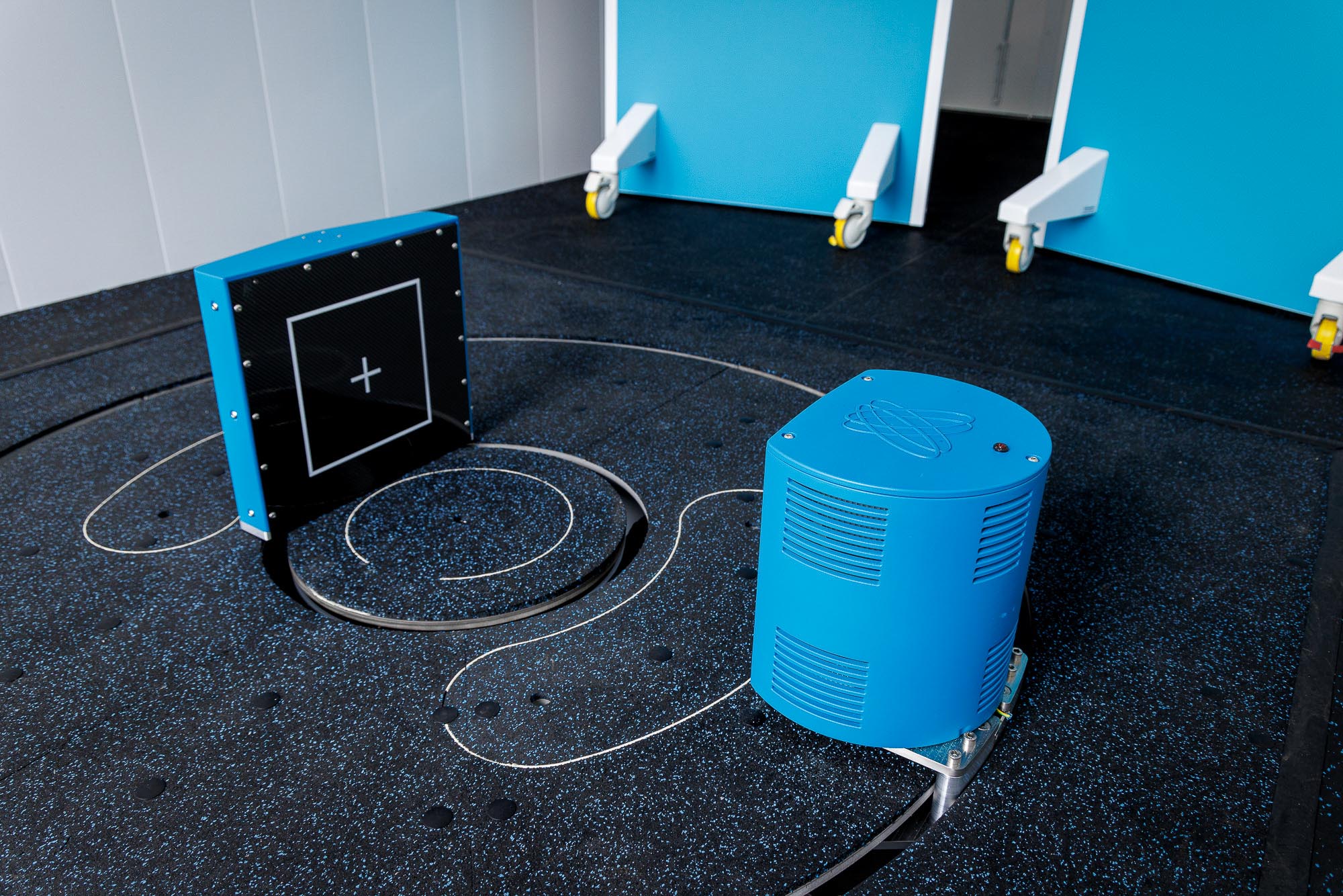
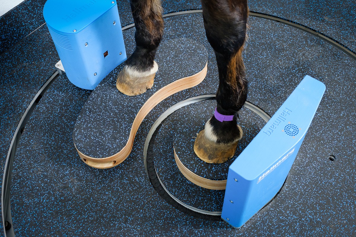
At the core of Vision CT’s design is Cone Beam (CBCT) CT technology, which differs significantly from traditional Fan Beam (FBCT) CT scanners. Cone Beam machines are noted for their open design, which includes a source and a detector but lacks the closed framework found in conventional CT scanners. Offering improved detail, CB technology is ideal for distal limb imaging, detecting non-displaced fractures, subtle changes in bone density, and small osteophytic lesions. The machine’s open architecture is especially beneficial for scanning horses, as it accommodates their size and allows for easy movement–-simply walking in and out of the scanning area without restriction. The open design also facilitates the standing position of the horse during scans, minimizing stress and eliminating the need for anesthesia or awkward positioning, thereby enhancing the safety and comfort of the animal.
Low Radiation and High Resolution:
An essential advantage of Vision CT’s CB technology is its low radiation levels, which are favorable compared to those of direct radiography (DR) systems commonly used in veterinary practices. With no general anesthesia required for the horse, and low-dose radiation allowing the operator and handler to remain in the room throughout, safety for all is assured. Despite the lower radiation dose, Vision CT achieves exceptionally high-resolution images, crucial for diagnosing intricate conditions in the equine limb. The novel dual-concentric design allows the patient to remain close to the detector for optimal image quality. This proximity of the horse’s leg to the detector, coupled with the machine’s ability to rotate 360° around the limb, ensures detailed, full-volume imaging of the targeted area.
Practical and Efficient Scanning Process:
Vision CT stands out with its practicality and efficiency. The system is designed for quick scans, completing a full rotation around the horse’s leg in just 60 seconds. This rapid scanning capability allows for a complete imaging procedure, from sedation to scan completion, in just minutes. Such efficiency is invaluable in a clinical setting, making Vision CT a highly practical tool for veterinary practices aiming to maintain high throughput without compromising on diagnostic quality.
Versatility in Application:
Vision CT’s design facilitates its use for the equine foot, pastern, and fetlock. Adjustments to accommodate different horse heights are made by using blocks for the horse to stand on, rather than altering the height of the device, further highlighting the system’s safe, horse-friendly design.
Complementary to Other Diagnostic Modalities:
Beyond its standalone benefits, Vision CT serves as a complementary tool to other imaging modalities such as MRI. Its ability to provide quick, high-resolution images of bony structures makes it an invaluable asset in cases where MRI provides soft tissue detail.
“Combining these advanced techniques has the potential to improve diagnostic sensitivity and specificity for these difficult injuries within the equine digital flexor tendon sheath.”
Dr Sarah Taylor (BVM&S MSc PhD Cert ES(Orth) DipECVS DipECVSMR MRCVS)
Senior Lecturer in Equine Orthopaedics, The Royal (Dick) School of Veterinary Studies, The University of Edinburgh
This synergy enhances the diagnostic capabilities of veterinary practices and creates a more comprehensive understanding of equine conditions. Also, Vision CT’s detailed 3D imaging capabilities can eliminate the need for more invasive diagnostic procedures, aid in surgical planning, and potentially reduce the overall need for surgery.
In summary, Vision CT is more than just another piece of advanced imaging equipment. By leveraging Cone Beam CT technology, Hallmarq has created a system that prioritizes the safety, comfort, and well-being of equine patients while delivering excellent diagnostic insights. This technology underscores Hallmarq’s role as a leader in veterinary imaging – paving the way for future advancements in the field.
Vision CT’s Flexible Installation Options and Future Directions in Equine Diagnostic Imaging
Vision CT by Hallmarq is not only a big leap forward in terms of diagnostic capabilities but also offers flexible installation options which cater to the diverse needs and constraints of veterinary practices. This means that a broad range of clinical settings can benefit from the advanced Cone Beam CT technology integrated within Vision CT. This wider-reaching accessibility reflects Hallmarq’s commitment to enhancing equine healthcare through innovative imaging solutions available to as many clinics as possible.
Flexible Installation Options:
The Vision CT system has been thoughtfully designed to accommodate two primary configurations, addressing various spatial and operational requirements. Firstly, the system can be seamlessly integrated into an existing room within a veterinary clinic. Thanks to its low X-ray power, which is effectively blocked by just 10cm (approximately 4″) of ordinary brickwork or blockwork, there’s no need for a specialized shielded room. This feature simplifies the installation process and reduces the infrastructure modifications needed for clinics to adopt this advanced technology. To facilitate easy access for equine patients, installation can include a raised floor allowing the horse to step up on a small ramp for scanning. It also incorporates protective screens for both the computer operator and horse handler––ensuring safety and ease of communication during the scanning process.
Hallmarq also offers an innovative, self-contained module configuration for Vision CT, resembling the size and shape of a shipping container. This modular approach provides unparalleled flexibility, allowing for the unit to be placed in a variety of locations, such as a parking area, without the need for extensive site preparation. The design of the module enables a straightforward walk-through process with horses entering from the front door and exiting through the back. And, this feature is particularly advantageous for scanning different parts of the horse without the need to reposition or turn the animal, which streamlines the workflow and enhances the overall efficiency of the diagnostic process.
“The installation was very straightforward. We opted for the Modular Room which meant the device arrived in its own purpose-built room. All we needed to do was lay a concrete pad. Once it arrived, it was ready to go. The horses are all very happy walking in and out of the low ramp and we have full ongoing support from the team at Hallmarq to ensure we are maximizing its use.”
Tom McParland, Surgeon, Valley Equine Hospital, UK.

Empowering Practices With Advanced Diagnostic Tools:
The flexible installation of the Vision CT system reflects Hallmarq’s vision for the future of equine diagnostic imaging – a future where advanced imaging technologies are accessible, practical, and seamlessly integrated into daily veterinary practice. By offering solutions that can be tailored to fit the specific needs and constraints of each clinic, Hallmarq is enabling a broader range of veterinary professionals to leverage the power of Cone Beam CT imaging. This democratization of advanced diagnostic tools is a significant step toward elevating the standard of care provided to equine patients worldwide.
Looking Ahead:
With its innovative design, fast scan times, safety for horse, handler, and operator, and flexible installation options, Vision CT is at the forefront of change within the field of equine diagnostic imaging. As veterinary medicine continues to evolve, embracing technologies like Vision CT will be crucial for practices aiming to stay ahead in the industry. The system not only enhances current diagnostic capabilities but prepares veterinary practices for future advancements, ensuring they remain equipped to provide the highest level of care. Hallmarq’s ongoing commitment to innovation, exemplified by Vision CT, promises to drive further advancements in veterinary imaging – improving diagnostic accuracy, animal health, and treatment outcomes.
In conclusion, Vision CT by Hallmarq represents real innovation in equine healthcare, promising a future where advanced diagnostic imaging is within reach for every equine practice. Its impact extends beyond the immediate benefits of improved diagnostic precision, heralding a new era of accessibility and efficiency in equine healthcare. As we look forward to other advancements on the horizon within the industry, Vision CT will undoubtedly play a pivotal role in shaping the landscape of veterinary medicine – fostering a deeper understanding of equine anatomy and pathology and improving the lives of horses and those dedicated to their care.
To find out more, please watch our video here.
[1]Morgan, Jessica M., Helen Aceto, Timothy Manzi, and Elizabeth J. Davidson. ‘Incidence and Risk Factors for Complications Associated with Equine General Anaesthesia for Elective magnetic resonance imaging’. Equine Veterinary Journal, 7 November 2023, evj.14026