


With a range of CT and MRI solutions for small animals and horses, we explore both advanced diagnostic imaging technologies in detail, the pros and cons of each, which one to use when and other key considerations.
When it comes to imaging techniques for patients at your veterinary practice, veterinary computed tomography (CT) and magnetic resonance imaging (MRI) are both viable options.
Much like human medicine, veterinary medicine has taken a giant leap forward with the widespread adoption of advanced imaging techniques in recent years. Computed Tomography (CT) and Magnetic Resonance Imaging (MRI) have revolutionized how veterinarians diagnose and treat animals, offering a window into the previously unimaginable body.
There are circumstances where the former is preferable, while other times, the latter is the obvious choice. That’s why it’s essential to understand each of them so you can be sure to offer the proper option.
Here’s a breakdown of the different use cases, benefits, advantages and disadvantages of veterinary CT vs. MRI for imaging your patients.
What You’re Imaging Determines Whether You Recommend Veterinary CT or Veterinary MRI
These two imaging solutions are optimized for different parts of the anatomy.
CT Scans in Veterinary Medicine
Computed Tomography, or CT for short, has become an indispensable veterinary diagnostic imaging modality in veterinary clinics and hospitals worldwide. At between 5-30 minutes for a scan, CT offers a quick way to acquire highly detailed images of the patient. The technology offers a remarkable blend of speed, detail, and versatility, making it invaluable for diagnosing a wide range of conditions. CT is primarily used for issues related to soft tissue, organs and muscles or bones.CT scans are painless and noninvasive and are generally recommended when traditional X-rays have been inconclusive.
At its core, a CT scanner uses X-rays, but is more powerful than a standard machine. Typically, your pet lies on a table that slides through a tube-shaped device, and X-ray beams rotate around its body, capturing images from multiple angles. A computer then combines these images to create cross-sectional 3D imaging, – allowing radiographers and veterinarians to examine each slice and indeed the patient’s internal anatomy in exquisite detail.
CT technology is advantageous in several scenarios. For instance, if a dog struggles to breathe, a CT scan can quickly reveal whether the issue stems from the lungs, heart, or surrounding structures. In cases of suspected bone tumors, CT not only shows the extent of the tumor but can also guide surgeons in surgical planning for the most effective treatment protocol.
CT scans have revolutionized veterinary care. They’ve helped make accurate diagnosis easier and surgeries more precise and have ultimately improved outcomes for animal patients. As technology advances, we can expect CT scans to become even more integral to veterinary medicine, offering ever-clearer windows into our pets’ health.
Magnetic Resonance Imaging Technology
Magnetic Resonance Imaging, or MRI, has transformed veterinary diagnostic imaging, particularly when examining soft tissues and neurological issues. MRI technology, which uses powerful magnets and radio waves instead of radiation, provides an unparalleled view of an animal’s body.
Imagine seeing inside your pet’s brain or spinal cord with incredible clarity without making an incision. That’s the power of MRI. It allows veterinarians to peer through skin, muscle, and bone to examine the delicate structures beneath.
When a pet undergoes an MRI, it lies still in a sizeable tube-like machine, so the pet is sedated. The MRI scanner creates a strong magnetic field around the animal, causing the hydrogen atoms in its body to align. Radio waves are then pulsed through the body, causing these atoms to produce faint signals. The scanner captures these signals and transforms them into detailed digital images by a computer.
The resulting high-resolution, detailed images are nothing short of remarkable. With the ability to review the patient in three dimensions (3D), they can reveal the tiniest abnormalities in the brain, pinpoint subtle changes in the spinal cord, or show the intricate structures within a joint. For a dog with unexplained seizures, an MRI might unveil a small brain tumor. A cat with a limp that appears normal on X-rays could identify a subtle ligament tear.
MRI has become an indispensable tool in veterinary medicine. It’s opened up new possibilities for diagnosis and treatment, allowing veterinarians to help treat those animals that might have been considered beyond aid just a few decades ago. As technology advances and becomes more accessible, we can look forward to MRI playing an even more significant role in keeping our pets healthy and happy
Comparing CT and MRI: When to Use Each
Veterinary CT is best for imaging bone and mineralized tissue. Therefore, it’s very effective for detecting skull abnormalities, spinal fractures, mineralized disc extrusion herniations and joint disease.
CT is also able to rapidly image a large area of the body while the patient is just sedated. Although MRI is superior for soft tissue imaging, it’s difficult to image the entire thorax and abdomen on MRI with the same rapidity as with a CT scan. For these tissues and areas, CT should be performed first.
This is particularly vital in emergencies where time is of the essence, such as checking for broken bones after an accident or assessing lung issues. CT scans use X-rays and computer processing to create detailed cross-sectional images, essentially providing a 3D view of the body’s internal structures compared to the 2D images obtained with radiographs.
MRI, on the other hand, is usually the imaging modality that is preferable for soft-tissue imaging and is excellent for depicting central nervous system (CNS) lesions. Unless the CNS disease is bone-related, CT likely won’t pick it up clearly, which can cause delays. For spinal cord imaging, veterinary CT often requires an iodine-based contrast dye to highlight the spinal cord, which can carry risk and add time to the process. In summary, veterinary MRI is the best option for soft tissue, muscles, the spinal cord, brain, ears and nose.
If your vet suspects a brain tumor, spinal cord injury, or soft tissue damage in a joint, MRI is likely to be their tool of choice. MRI uses powerful magnets and radio waves to produce detailed cross-sectional images, offering a comprehensive 3D view of soft tissues.
In some complex cases, both CT and MRI might be used to get a complete picture. For instance, in cancer cases, a CT might check for the spread to the lungs or bones, while an MRI could provide detailed images of the primary tumor.
| Feature | CT (Computed Tomography) | MRI (Magnetic Resonance Imaging) |
| Technology | X-rays | Magnetic fields and radio waves |
| Best for | Bone, chest and abdomen emergency cases | Soft tissues such as muscles, neurological issues |
| Scan Time | Minutes | 40-45 minutes |
| Radiation | Uses ionising radiation | No ionising radiation |
| Detail | Excellent for bone and lung | Superior for soft tissue |
| Cost | Generally lower | Generally higher |
| Availability | More common | Less common |
| Anesthesia | Often required, shorter duration | Almost always required, longer duration |
| Contraindications | Few (pregnancy) | Metal implants, some medical devices, in addition to the requirement for general anesthesia which may be contraindicated in an individual patient |
MRI Provides Higher Quality Images to Support Definitive Diagnoses
Both veterinary CT and MRI can be used for diagnosis, but each has specific advantages for certain use cases. Soft tissue and muscle imaging on MRI, for example, are so well defined that a problem’s severity can be observed, which is rarely the case when using CT. For example, it enables you to identify the margins of a lesion prior to surgery. You can determine the size of an individual lesion in the brain and spinal cord, even those as small as 1-2mm.
With veterinary CT, you might see a reasonably sized mass, but MRI allows you to identify smaller lesions, such as hemorrhage. The detail that MRI provides regarding nervous system anatomy allows a precise determination of the structures affected (such as white matter, gray matter, cranial nerves, etc.), which is essential to more accurately consider the responsible disease.
Beyond showing the extent of the primary disease, MRIs can reveal the impact it has on the tissue surrounding it. For example, you might see a mass or lesion in the brain and diagnose it as a tumour, and you might also observe oedema as a reaction to it. Seeing the secondary consequences of the disease provides direction for a clear treatment plan and your prescription can address all the complications the patient is experiencing. Whereas CT too often lacks this level of detail. Instead, everything merges into a single abnormality.
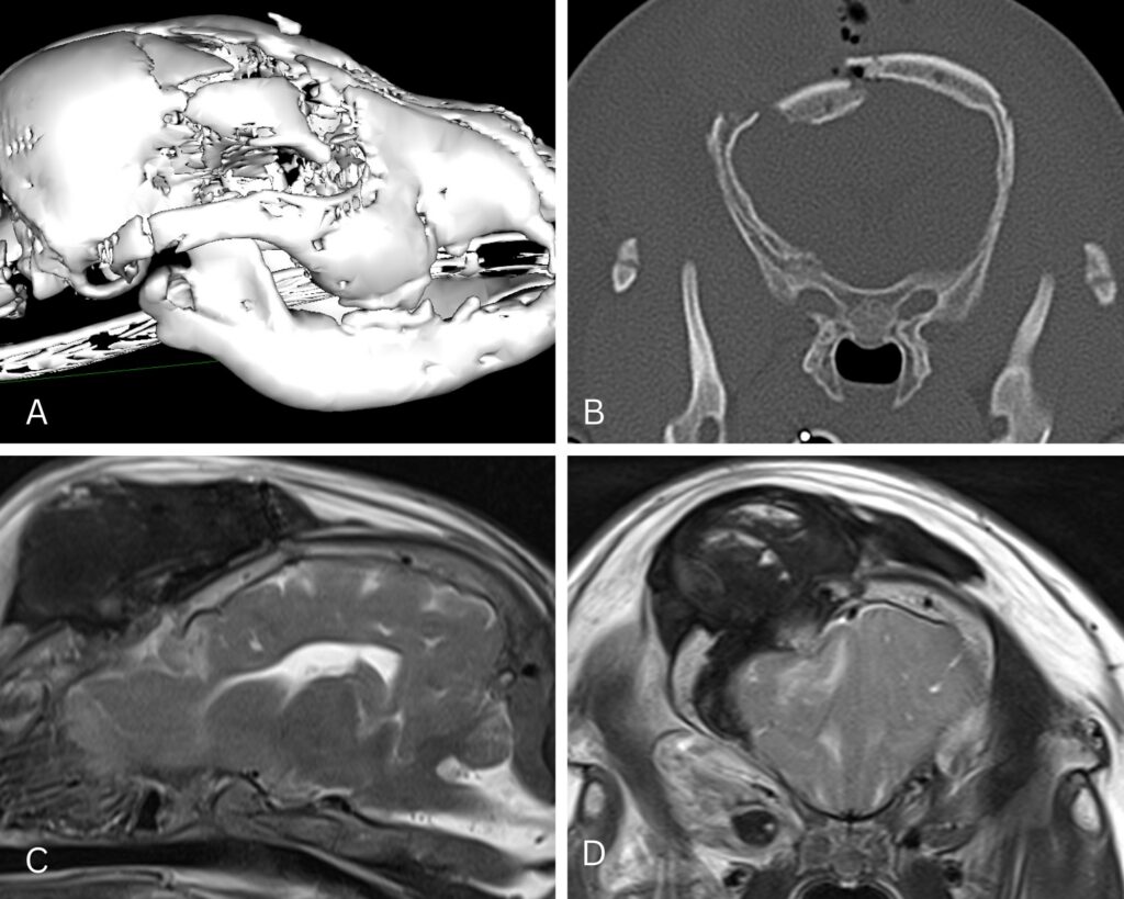
In Fig. 1 (above): Clinical examination suggested that this 3-year-old cat diagnosed with a dilated pupil (mydriasis), was not suffering from a disease affecting the eye itself, but that the condition was related to a disease of the brain. A rapid understanding of the cause can be identified with MRI to avoid unnecessary treatment attempts and improve the situation with early medical intervention.
With a CT scan, the use of X-rays means there’s some radiation exposure, although the risk is generally considered low. Additionally, CT excels at imaging bones and lungs but is less effective for detailed views of soft tissues like the brain or spinal cord. Types of tumours can be defined more clearly with MRI, especially using contrast. This helps you arrive at a better diagnosis so you can create a more targeted treatment plan.
Some Patients Aren’t Good Candidates for MRIs
Having explained the benefits of MRIs, it’s time to address occasions when they are not a viable option. For example, if a patient has any metal in their body (say, from surgical implants or identification microchips), it can distort the MR imaging. The metal may be far enough away from the scan for it not to be a factor: a hip implant won’t impact a brain scan, for example. It would, however, distort a pelvic scan. Even a pet’s microchip can interfere with image interpretation of surrounding tissues, so it is vital to check with the MRI referral centre when considering the animal’s individual circumstances. A distorted image is of limited value in diagnosing patients. It should be noted that pets with pacemakers can’t be MR imaged at all since the magnet could interfere with the device.
There are also other potential drawbacks to MRIs, which will be explained below.
Additional Benefits of CT to Consider
Unlike CTs, MRIs require anesthesia to keep the patient still during the study, which could be deemed a health risk for some pets. Accordingly, older pets or ones with existing health complications may require additional testing to see if MRI is a good fit for them. The tests to ensure that anesthesia is safe for the patient can add time, drive up the cost and generally frustrate clients.
Another advantage CT has over MRI is speed. MRI takes 40-45 minutes, while CT can produce images in just 10 minutes, and a CT can image several regions such as thorax and abdomen during this time. This makes CTs far more manageable for traumatised patients.
MRI is generally more expensive than other diagnostic imaging tests. It’s also usually easier to get a CT. There can be a waiting list for MRIs since the length of the procedure means fewer can be administered each day. For emergency cases, in particular, veterinary CT is generally more available than an MRI. For many pet owners, these challenges are not deemed insurmountable when the health of their cat or dog is a priority.
A Pet Owner’s Perspective: Preparation for Veterinary Scans
When your veterinarian recommends a diagnostic imaging procedure scan for your pet, it’s natural to feel a mix of hope for answers and concern about the procedure. Understanding what to expect can help ease those worries and ensure the best possible experience for you and your pet.
Use of Sedation and Anesthesia
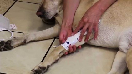
CT scans generally need lighter sedation since the procedure is quicker. MRI scans take longer and require general anesthesia to keep the patient still. This helps manage the animal’s stress or anxiety, especially in a noisy environment like an MRI machine. There is also additional padding to ensure the animal is appropriately positioned.
While most pets tolerate sedation well, your veterinarian must conduct a thorough pre-scan evaluation to assess potential risks based on your pet’s health history. The type of sedation or anesthesia used will depend on the specific scan and your pet’s needs.
Interpreting Results
Once the CT or MRI scan is complete, the next step is interpreting the results. This process is far more complex than simply looking at pictures. Accurately reading these sophisticated clinical images requires years of specialized training and experience.
Typically, the scans are analyzed by a veterinary radiologist, a specialist dedicated to understanding and interpreting resolution images. They scrutinize every detail, comparing what they see to what’s considered normal and looking for patterns and abnormalities that might indicate specific diseases or conditions.
For a CT scan, the radiologist might examine the density of different tissues, looking for unusually light or dark areas. They’ll also check the size and shape of organs, the bone structure, and any masses or abnormalities. For an MRI scan, the radiologist will often look at the intensity of signals from different tissues, which can reveal subtle changes in soft tissue injuries.
The radiologist will compile their findings into a detailed report. This document describes what was observed in the images, often using medical terminology that might seem foreign to most pet owners. The radiologist might also include their interpretation of these findings, suggesting possible diagnoses or recommending further tests if needed.
This report is sent to your veterinarian who will translate these complex findings into actionable information. Your veterinarian will consider the radiologist’s report in the broader context of your pet’s symptoms, physical exam findings, and other test results. They might consult with the radiologist or other specialists to completely understand what the images reveal.
Long-Term Treatment
Long-term care and follow-ups are crucial for animals recovering from health risks. Ongoing monitoring and support help prevent complications and ensure the best possible recovery. Specific recommendations for follow-up appointments, rehabilitation services, and patient education are vital to ensuring adherence to treatment plans.
These follow-ups might include additional diagnostic imaging tests, physical therapy, or medication adjustments. The goal is to ensure your pet continues to recover well and address any issues that may arise during the recovery process.
Summary
The fact is that veterinary CT and MRI should be seen as complementary. During an animal’s lifetime, as one of your patients, they may require a CT and an MRI for different conditions.
Frequently Asked Questions (FAQs)
Is CT or MRI Safer for my Pet?
Both diagnostic recommendations are generally safe when performed correctly. The main risk in both procedures comes from the anesthesia not the scanning itself. MRI doesn’t use radiation, which some consider an advantage, but the slightly higher radiation exposure from a CT scan is typically not a significant concern for pets. Your veterinarianwill recommend the safest and most appropriate option based on your pet’s specific medical condition. The veterinarian will make sure the pet is in a comfortable position when performing diagnostic imaging procedures.
How Long Must my Pet Stay at the Clinic for a CT or MRI Scan?
Most pets will be seen on an outpatient basis and can go home on the same day as their scan. The entire process, including preparation, the scan itself, and recovery from anesthesia, usually takes 2-4 hours for a CT scan and 3-6 hours for an MRI. Your veterinarian will give you a more precise estimate based on your pet’s needs.
Will my Pet Feel any Pain During or After the Scan?
The scans themselves are painless. Your pet will be under anesthesia during the procedure and shouldn’t experience any discomfort. Some pets might feel dizzy or disoriented as they recover from anesthesia, but this typically passes quickly.
Can I Stay With my Pet During the Scan?
For safety reasons, pet owners are not allowed in the room during the scan. The machines use strong magnetic fields (MRI) or levels of radiation (CT), which can be harmful to humans with prolonged exposure. Rest assured, though, that trained professionals will continuously monitor your pet throughout the procedure.
How Soon Will I Get the Results?
Full results typically take 24-48 hours, as a specialist must interpret the images carefully. In emergencies, preliminary results may be available sooner. Your veterinarian will contact you to discuss the findings as soon as they’re available.
What if my Pet is too Large for the MRI Machine?
Size can sometimes be a challenge, especially for very large dogs. Some veterinary hospitals have open MRI machines that can accommodate larger pets. If this isn’t available, your veterinarian might recommend alternative imaging methods or refer you to a facility with suitable equipment.
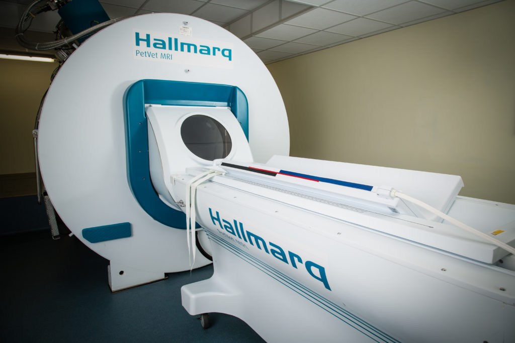
For example, the Hallmarq zero-helium, small animal, 1.5T MRI machine is designed specifically for pets, not people! Its V-shaped patient bed ensures comfort for the smallest of Chihuahuas or the very largest of Dobermans or Great Danes. At the same time, its dual coil function enables faster scanning of the head and spine, making for a better overall experience for your pet.

Are There Alternatives to CT and MRI Scans?
Yes, there are alternative diagnostic tools depending on the medical condition being investigated. Alternatives might include X-rays, ultrasounds, or nuclear medicine scans. Your veterinarian will recommend the most appropriate imaging method for your pet’s specific needs.
Remember, your veterinary team is your best resource for information about your pet’s specific situation. Don’t hesitate to ask them any questions you might have about imaging options or any other aspect of your pet’s care.
Authored by Dr Simon Platt, Small Animal Medical Director with Hallmarq Veterinary Imaging
INTERESTED IN VISIONARY VETERINARY IMAGING?