

A recent review of over 100 papers on advanced imaging summarised just 20 to showcase major technological developments in equine imaging across all modalities. Guest blogger, Fiona Farmer, takes a closer look at their findings.
Advancing access for equine welfare
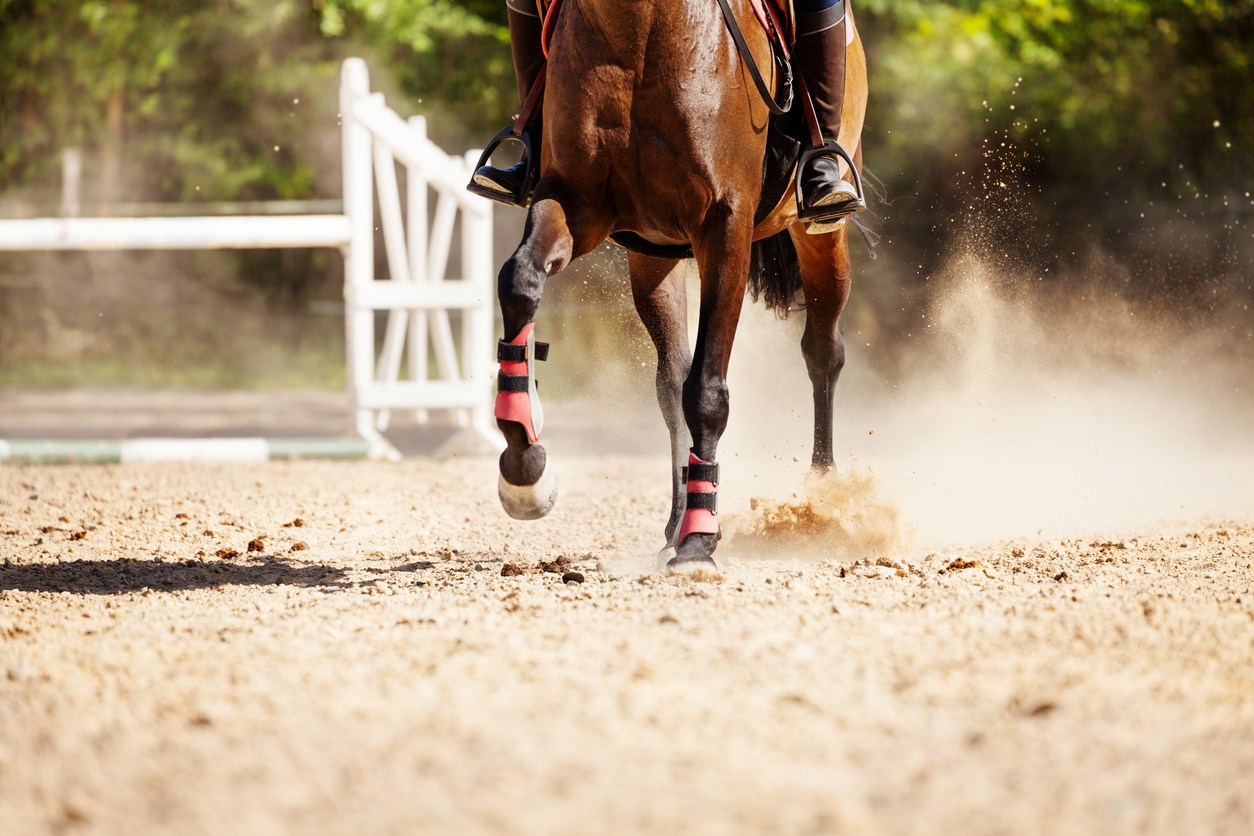
Lameness is one of the most common health and welfare concerns for horses. Injuries or diseases of the distal limb make up the majority of these cases.
“The centuries-old adage “no foot, no horse” still holds true today, with the foot the main focus of much imaging.”
DR (digital radiography) technology is now over 100 years old but still remains the most commonly used imaging technique. However, recent advances in diagnostic imaging, including CT (computed tomography) and MRI (magnetic resonance imaging), along with an ever-increasing growth in knowledge, has dramatically improved equine welfare and safety.
Recognition of these advances was recently highlighted by The British Equine Veterinary Association (BEVA). Veterinary Radiology & Ultrasound (VRU), Equine Veterinary Education (EVE) and the Equine Veterinary Journal (EVJ) showcased major technological developments in imaging across all modalities. Lameness experts Mathieu Spriet, Ann Carstens and Tim Mair, jointly picked 20 out of almost 100 available papers. They then concisely summarised the advances in MRI, CT, Ultrasound, PET, and radiography.
The virtual edition, available to everyone until mid-April, is available here
Focus on the foot
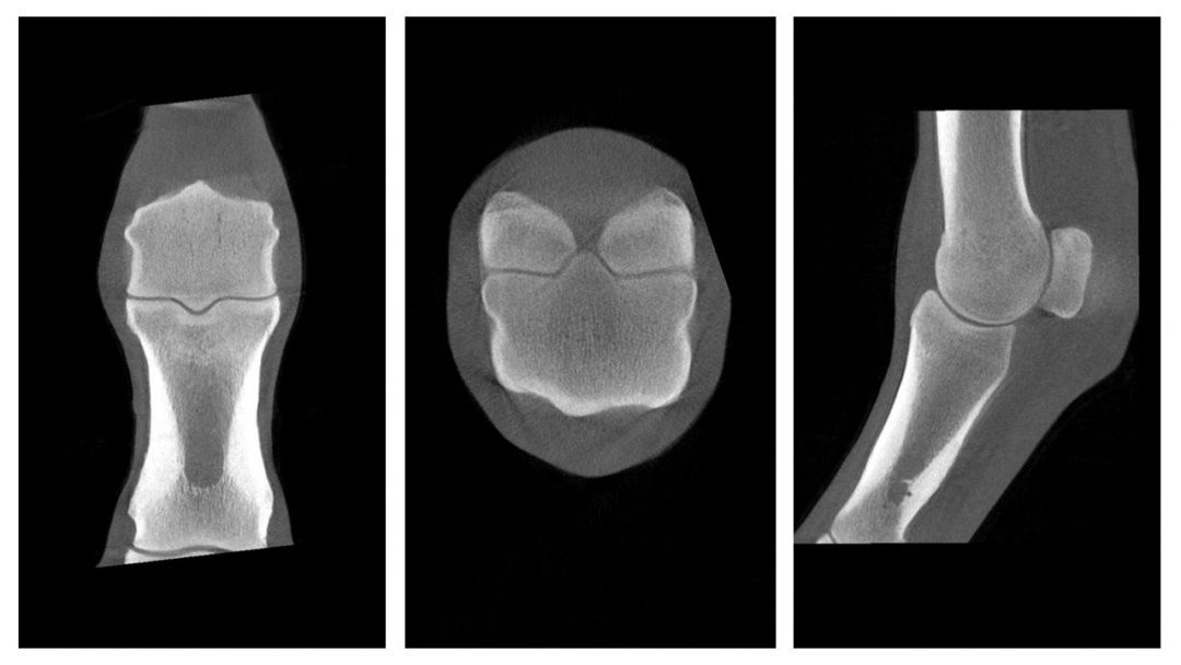
Of those papers selected, six relate to CT and six to MRI. Mathieu Spriet composed a wonderful overview in which he noted that the foot (and fetlock) remains a firm focus of advancing imaging. One of the first radiographs taken in 1895 was of the foot, and as CT and MRI continued to develop in the early 2000s, it was once again the foot that was the area of interest.
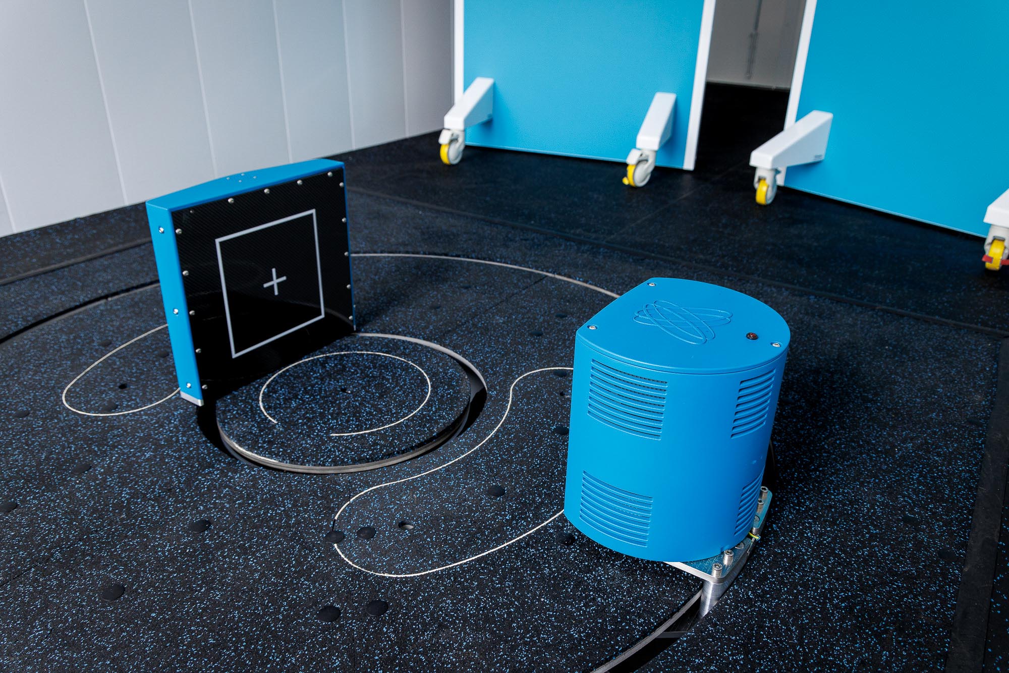
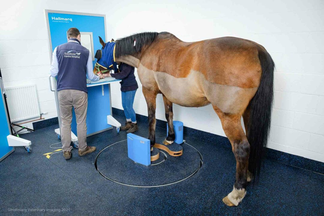
Hallmarq is proud to be pushing the boundaries of what can be achieved with advanced diagnostic imaging. Our Standing Equine leg CT machine enables imaging of the distal limb under standing sedation, thus removing the need for general anaesthesia. This has improved safety for horses, made advanced imaging more affordable and accessible to owners, and increased the number of horses that can undergo advanced imaging. All this combines to increase patient welfare and veterinary knowledge.
Technological advances in CT
So, how have technological advances in CT enabled veterinary progression? Undoubtedly removing the requirement for a general anaesthetic has been, as Spriet described, a revolution. Standing CT utilises cone beam technology to rotate around the area of interest and provide a cross-sectional image of the distal limb, negating the need for the horse to be fully anaesthetised. The benefits of this include:
- Safer for horse and handler – lower dose of radiation, and no need for a general anaesthetic.
- Affordable – minimal installation and running costs and no need for a purpose-built room.
- Effective – spatial resolution locates small changes in the distal limb, with no superimposition or complex overlap of anatomy.
- Quick image acquisition time – 3D image sets can be captured in 60 seconds with the whole procedure taking just 15 minutes.
Not forgetting MRI
The use of MRI in orthopaedic imaging has been a reliable modality for many years. A review of the papers shows that there have not been any major technological advancements in recent years. However, MRI continues to be an improving modality due to increased knowledge and image acquisition techniques. Many studies using MRI have been used to explore and identify the pathophysiology of numerous conditions including condylar fractures, subchondral defects, sagittal groove injuries of the proximal phalanx and collateral ligament desmitis of the distal interphalangeal joint.
Significant advances in imaging of the distal limb
The virtual edition includes a collection of papers on scintigraphy, ultrasonography and radiography. Ultrasound continues to improve steadily and one paper explores non-weight-bearing imaging. Although papers on radiography are not frequently published today, this imaging modality remains popular among equine clinicians. Even after 100 years, the technique continues to improve for image acquisition and diagnosis. Likewise, scintigraphy use is declining but remains a useful tool in some situations.
This review, highlighting some of the most significant advances in distal limb imaging from the past five years, is free to access until mid-April and can be found here
If you would like to discuss advanced imaging with our team, we welcome you to contact us
Fiona Farmer is an FEI 3* official vet, part-time content writer with Companion Consultancy, busy mum, horse owner and rider, and fitness class founder.