


Ever wondered what the difference is between a low-field and a high-field MRI? Hallmarq Small Animal Advisory Board member Dr. Jennifer Brisson sheds more light on the question that will, at some point, have crossed the mind of anyone employed in the veterinary field.
Let’s Talk Tesla
The definition of low-field vs high-field MRI is determined by magnet field strength. Generally speaking, low-field MRIs are less than one Tesla (T) whereas high-field MRIs are 1.0T or greater. There are pros and cons to each type of MRI and understanding the difference is important when making a selection for your patient or practice.
The Right Tool for the Job
So, why is magnetic field strength important? Magnetic field strength determines the MRI signal, which, in turn, informs the patient’s image. In more specific terms, the MRI signal is proportional to the square of the main magnetic field strength. Thus, MRI signal, and signal-to-noise ratio (SNR), increases with increasing field strength. Higher signal quality leads to better image resolution. So then, why not just use high-field MRIs every time? Let’s review:
Low-Field MRI
Historically, low-field MRIs are resistive or permanent type magnets. They come in lower field strengths 0.3-0.5T, which generate less signal when compared to high field strength units. With less signal comes reduced spatial resolution and longer scan times. There may also be limitations in pulse sequences, including chemical shift fat suppression and MR spectroscopy. Low-field MRIs are generally smaller, less expensive to install, and require less shielding. However, daily power usage of resistive-type scanners can be quite high thus increasing operating expenses.
Low-field MRIs usually do not require helium. One distinct advantage of low-field MRIs is the open design which improves patient access and visibility. Let’s take a look at the advantages vs disadvantages:
- Advantages: open design with large bore diameter, less expensive to purchase and install than high-field MRIs, requires less shielding, no helium requirement
- Disadvantages: Lower image resolution, increased scan times, limited pulse sequences
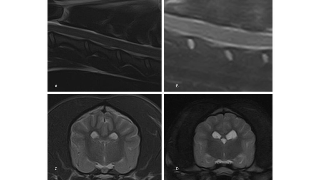
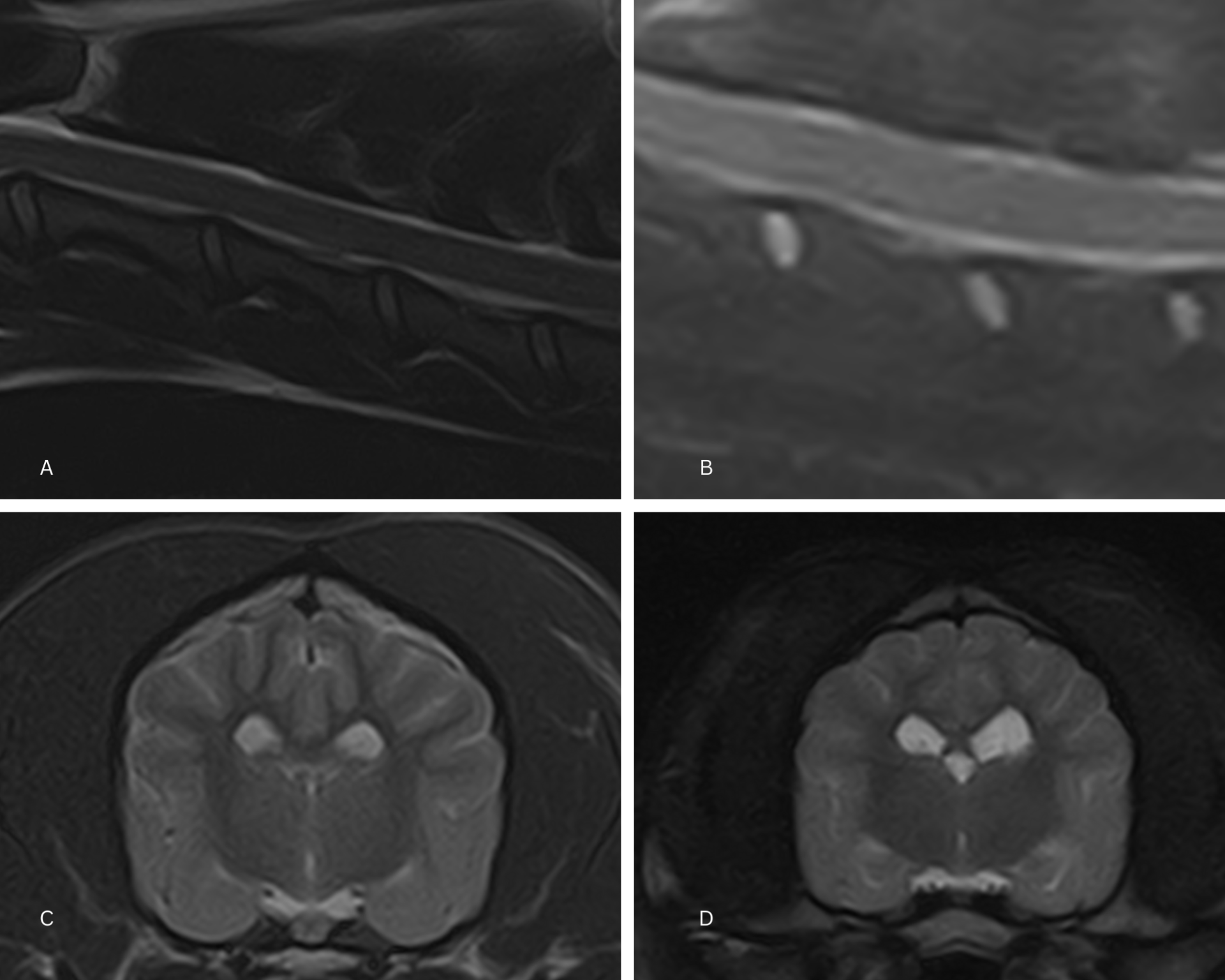
Figure 1 – A normal cervical vertebral column can be seen on the top of the image panel with a high-field (A) and low-field version (B). A normal brain can be seen on the bottom panel of the image with a high-field (C) and a low-field version (D).
High-Field MRI
High-field MRIs are superconducting magnets, and generally range from 1.5-3.0T for most clinical applications. With the higher field strength comes a greater SNR. The higher signal greatly improves spatial, contrast, and temporal resolution when compared with low-field strength units. Pulse sequence availability is broad and constantly improving. Historically, these units have relied upon helium for operation which can be costly and challenging to obtain. However, new designs have greatly reduced or eliminated helium requirements for 1.5T units, reducing the cost of ownership and improving sustainability.
With higher field strengths, greater shielding is usually required and increases install expense. High-field MRIs typically utilize a closed design that can limit patient size, access, and comfortability. Let’s weigh up the pros and cons:
- Advantages: Superior SNR, superior image resolution, wide sequence capability, shorter scan times
- Disadvantages: expense, closed design, varying helium requirements
Informed Choices
In conclusion, MRI is one of the most effective noninvasive diagnostic tools available in medicine and can bring great value to patient care. Both low-field and high-field MRIs are in common use in veterinary practice, and veterinary-specific MRI units are available in both categories. Understanding the advantages and disadvantages of low-field vs high-field MRIs will allow for informed decision-making when selecting for the individual patient or practice.
INTERESTED IN VISIONARY VETERINARY IMAGING?