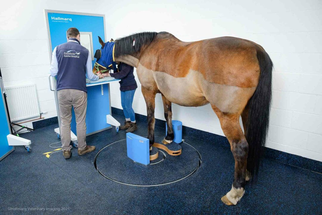
Handling the complexities of equine lameness, especially when it displays in the foot and fetlock, is much like trying to solve an intricate puzzle. Surprisingly, given these inherent difficulties, around a third of all equine veterinary visits are focused on lameness issues. Second only to skin conditions, they are reported as being the most common problem affecting horses1. This statistic emphasizes the critical need for precision in lameness diagnosis and treatment. Here’s where Vision CT by Hallmarq emerges as the unique solution to redefine the standards of equine care.
Bridging the Diagnostic Gap in Equine Lameness
The Diagnostic Dilemma
Lameness in the foot and fetlock presents a complex challenge. Traditional imaging techniques, while necessary, often provide incomplete pictures, leaving veterinarians seeking more detailed insights for their diagnoses. Thankfully, Vision CT can provide unprecedented precision and clarity by harnessing the power of Cone Beam CT – technology optimized for the distal limb.
A Leap Forward with Cone Beam Technology
Distinguished by its sub-millimeter resolution and ability to perform detailed 3D reconstruction, the Vision CT system brings into focus the intricate bony pathology often missed by other modalities. This enhancement is crucial for cases where a primary bone injury is suspected, significantly aiding in surgical planning, and offering invaluable insights into the articular surface of joints.
“Contrast arthrograms have been to assess the distal interphalangeal and metacarpo/tarsophalangeal joint for potential articular cartilage loss. Vision CT has been useful for cases with sagittal groove trauma of the proximal first phalanx, keratomas, and other foot problems.”
Dr Ellen Singer BA, DVM, DVSc., Diplomate ACVS & ECVS, FRCVS, American, European and RCVS Specialist in Equine Surgery, Veterinary Associate, Sussex Equine Hospital, UK
The Vision CT advantage: Precision, Non-Invasiveness, and Efficiency
Transforming Equine Imaging
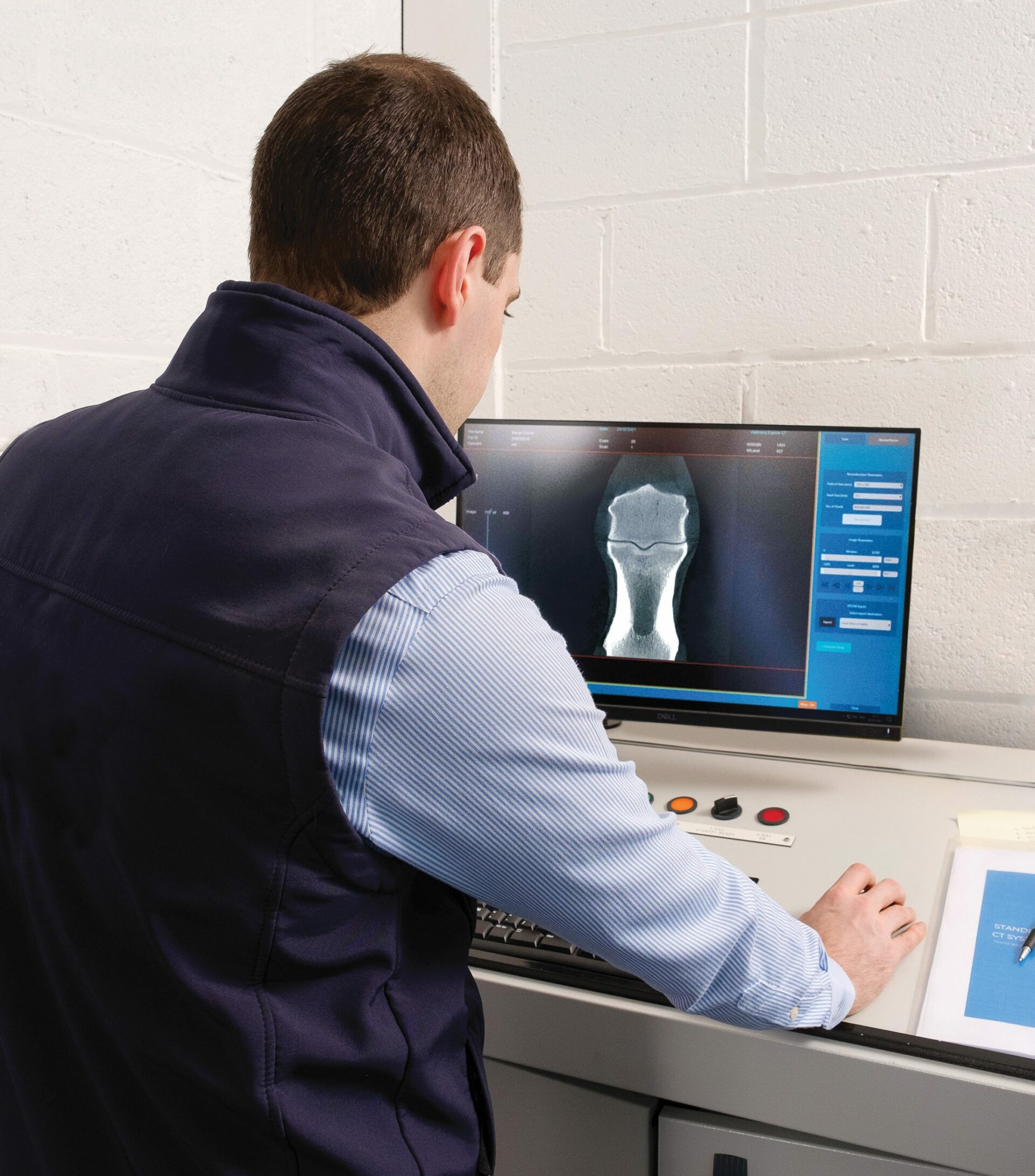
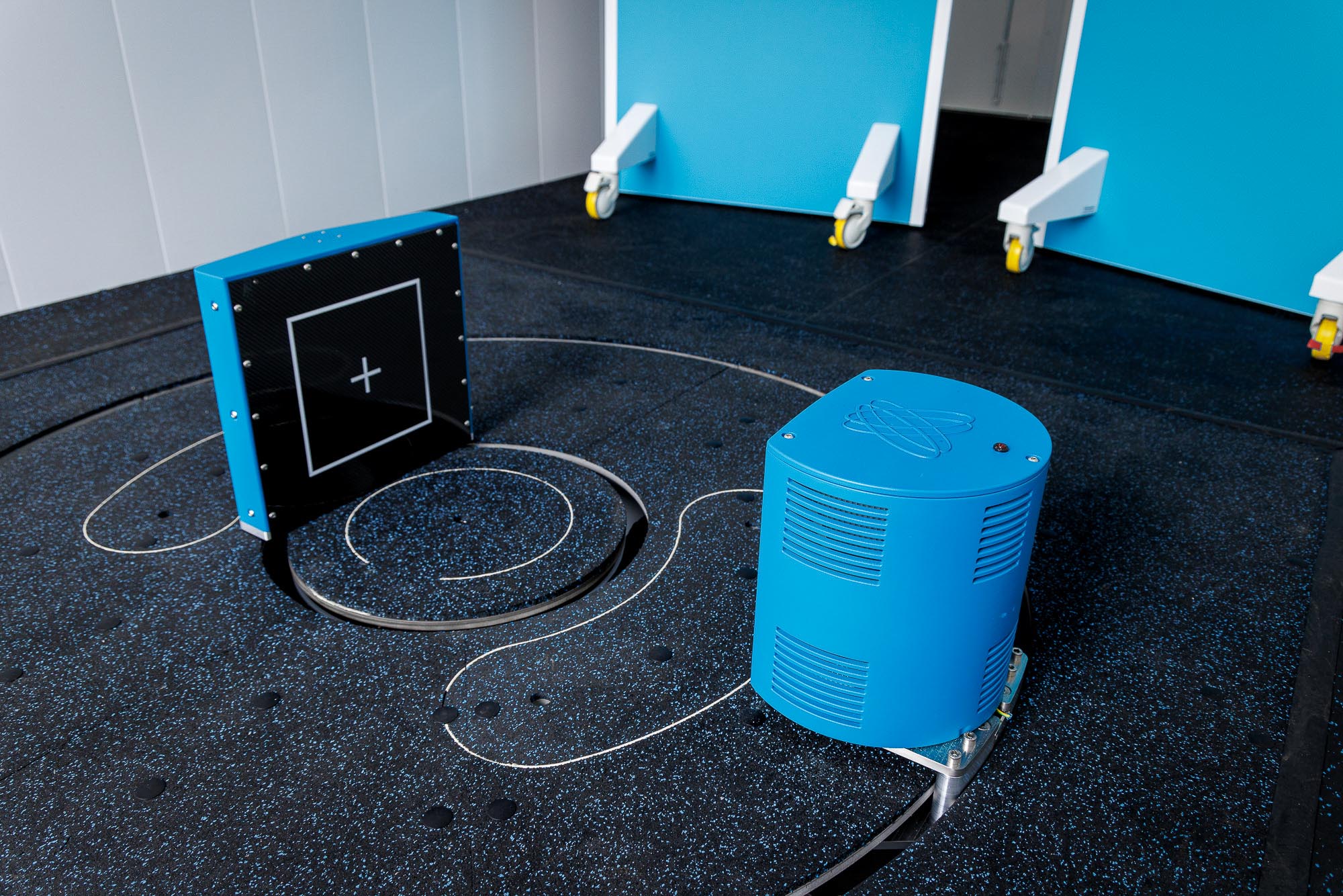
Vision CT stands out with its non-invasive approach, low radiation levels, and capacity to capture scans on the standing, sedated horse. This setup, noticeably different from conventional CT imaging, allows for a comprehensive 3D picture of the limb to be constructed in just 60 seconds; a particularly beneficial feature, since general anesthesia is avoided.
Enhancing Surgical Outcomes and Treatment Plans
The detailed 3D imaging provided by Vision CT facilitates precise surgical planning. In addition, it enables a complete evaluation of small bony injuries that might otherwise be missed. For surgical cases, especially those involving the distal limb, access to such detailed pre-operative imagery ensures interventions are as targeted and effective as possible.
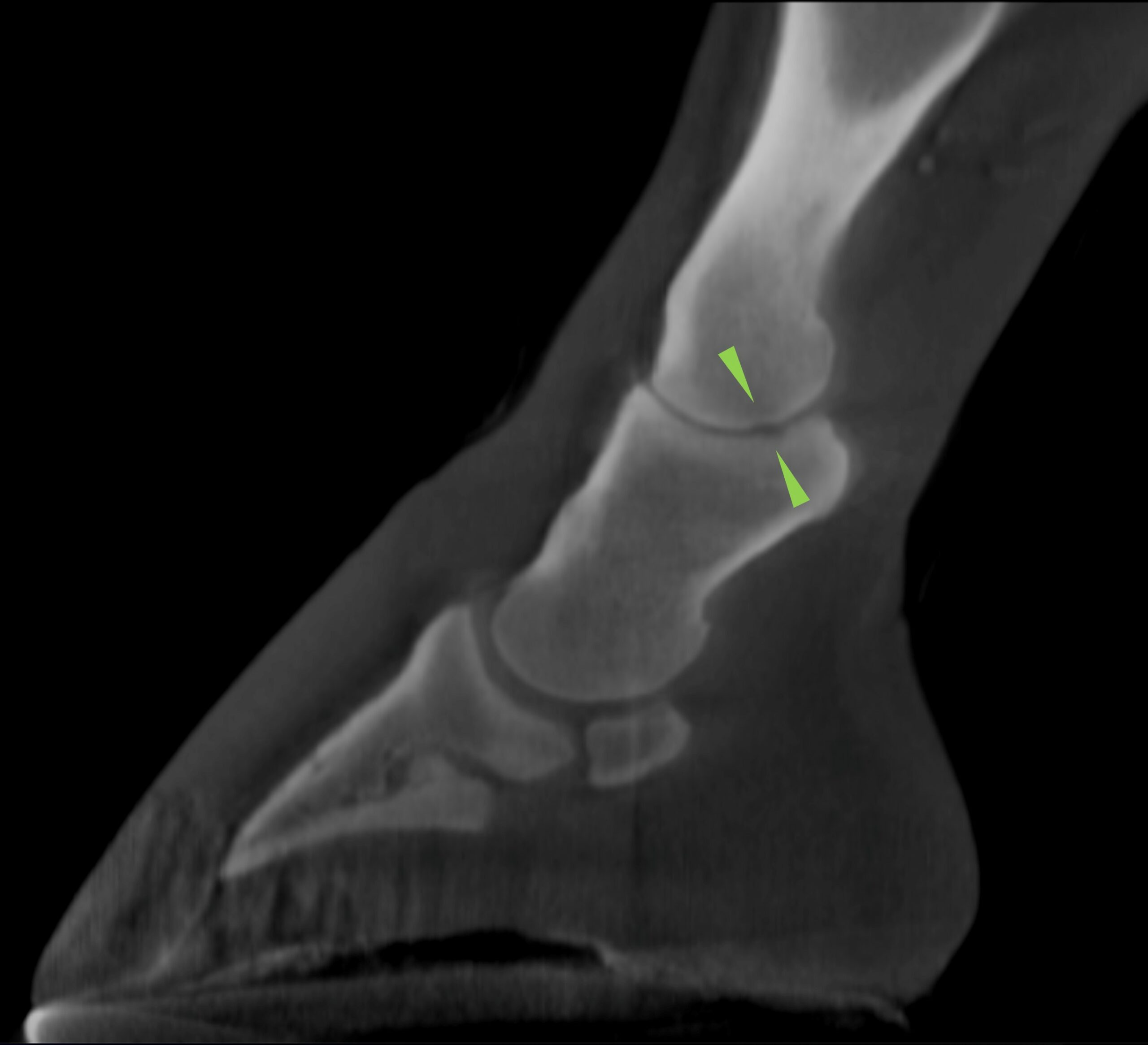
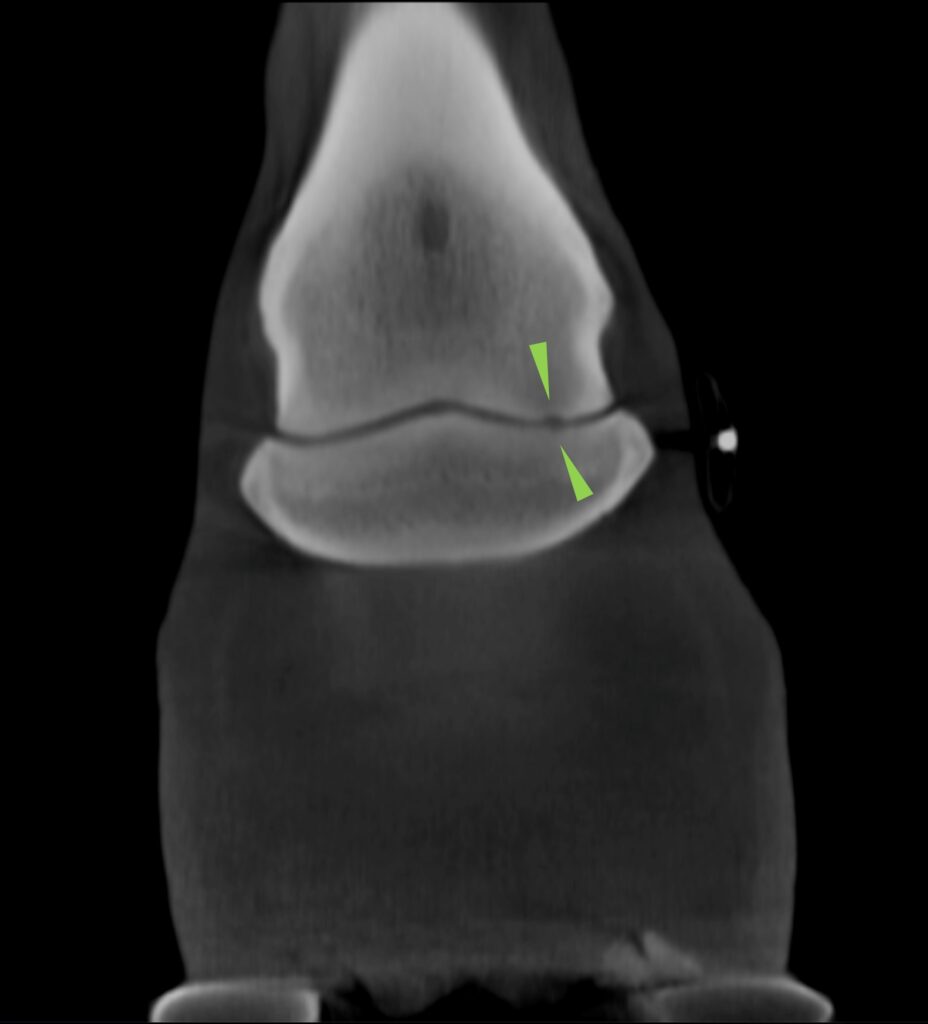
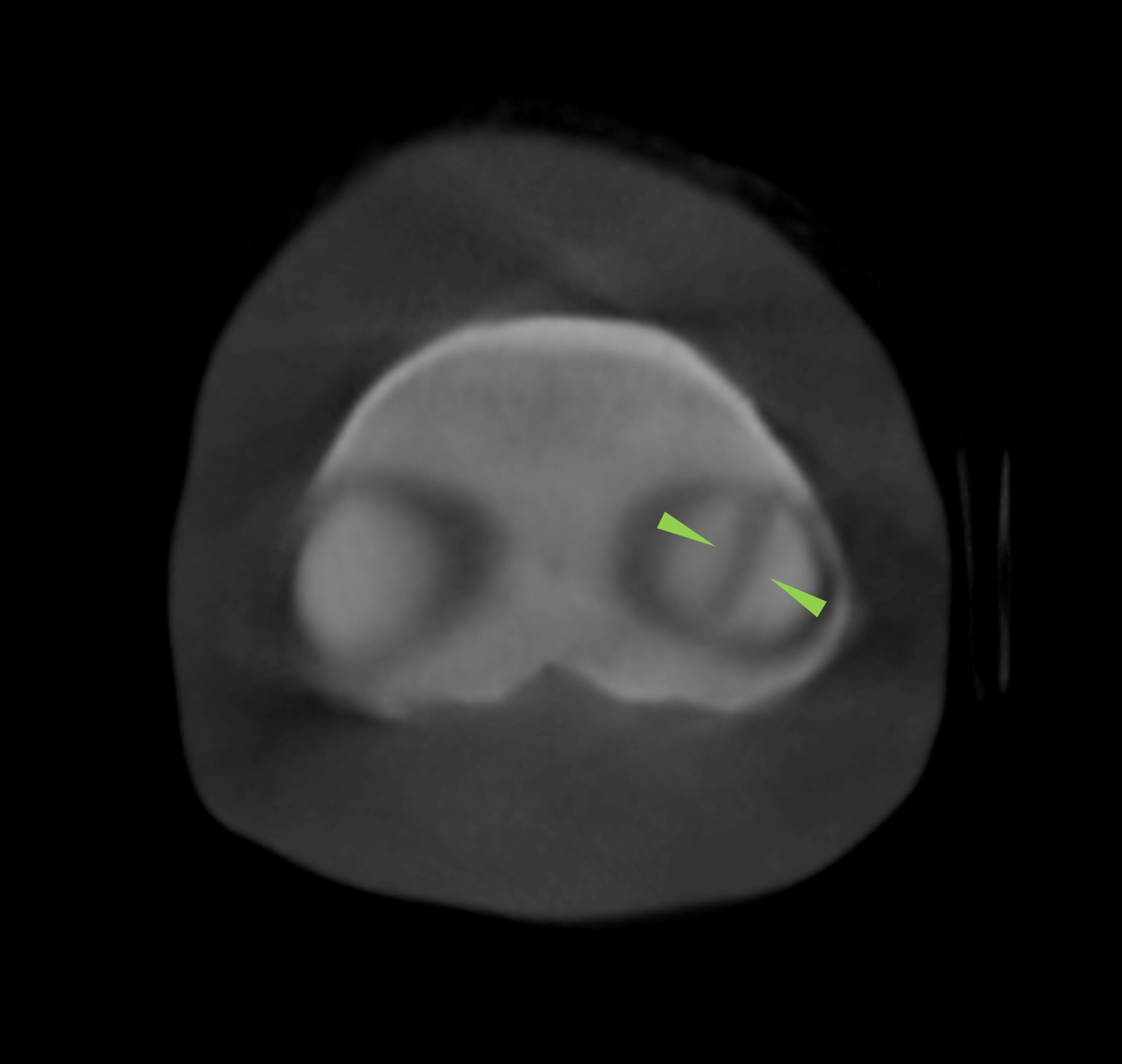
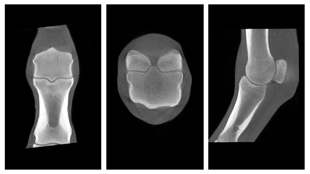
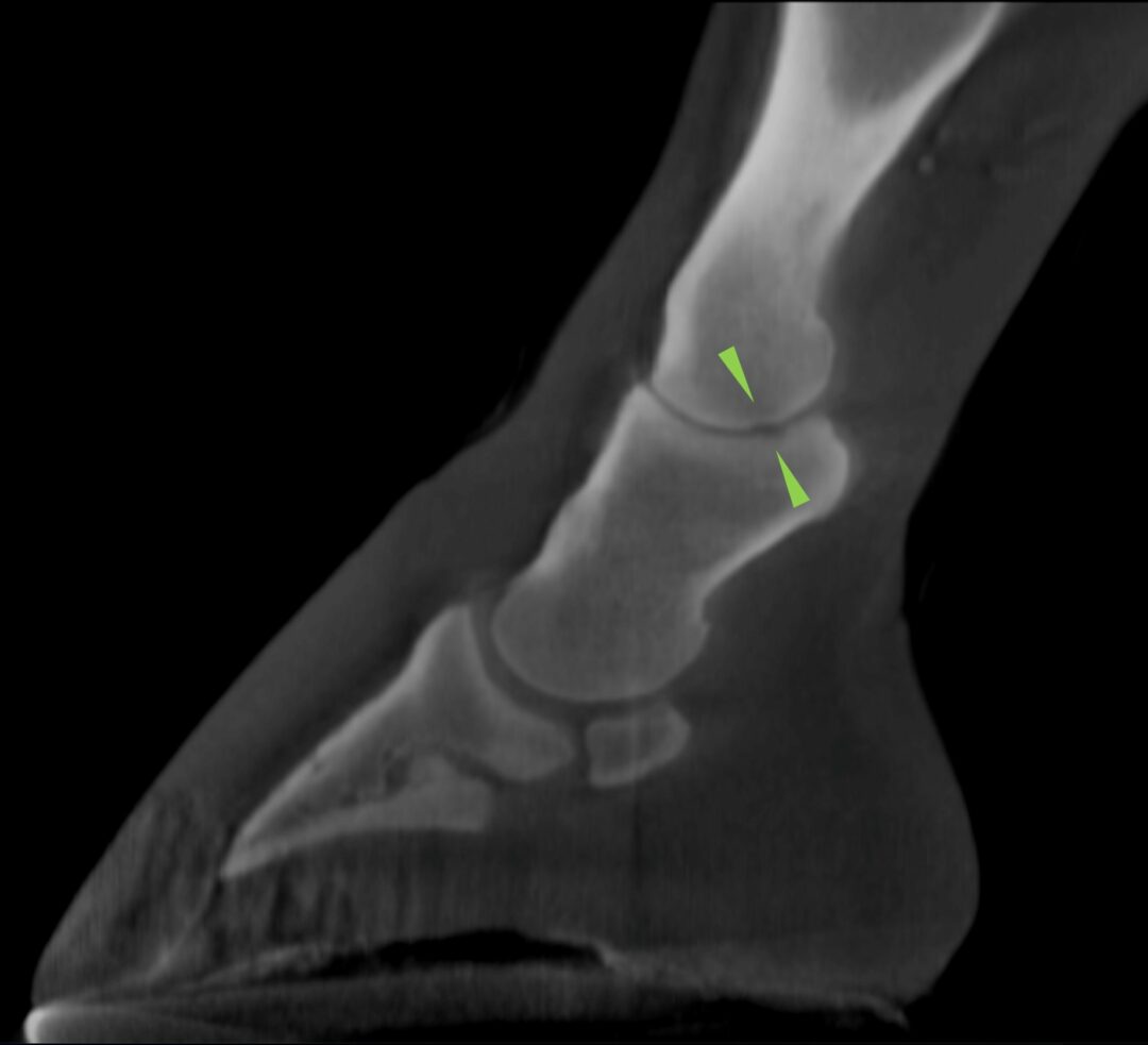
Figures 1, 2, and 3 (above) show that a small and poorly marginated region of hypoattenuation of the adjacent trabecular bone is present. A shallower kissing lesion extends in the same direction through the subchondral bone plate of the lateral glenoid of the middle phalanx with no extension to the adjacent trabecular bone.
Expanding the Horizon of Equine Care with Vision CT
3D Musculoskeletal Imaging at Your Fingertips
For clinics with a significant sports medicine caseload, Vision CT introduces an era where 3D musculoskeletal imaging becomes a tangible reality. This advancement is invaluable, offering a clear picture of everyday lameness and empowering veterinary professionals to make informed decisions with confidence.
Tailored Imaging For Comprehensive Care
Beyond its diagnostic facilities, Vision CT’s ability to scan multiple regions of the distal limb (coupled with its superior bony tissue differentiation) changes what is possible in equine healthcare. Whether diagnosing subtle changes in bone density, detecting non-displaced fractures, or differentiating between sub-chondral and cortical bone pathology, the Vision CT system provides a transformative level of detail.
In the ever-evolving landscape of equine healthcare, staying abreast of technological advancements is paramount. Accordingly, Vision CT is at the forefront of this innovation, offering solutions that combine efficiency, precision, and safety. Its integral role in helping diagnose and manage lameness, particularly in the foot and fetlock, heralds a new era in veterinary care.
Embrace Advanced Diagnostics with Vision CT
The journey towards excellence in equine diagnostics is paved with innovation. Vision CT stands ready to transform your approach, setting new benchmarks in patient care. Whether for routine evaluations, complex surgical planning, or comprehensive sports medicine caseloads––its potential impact is undeniable.
“”Hallmarq’s Vision CT has delivered 3D images of feet, pasterns and fetlocks in standing horses in a practical, quick and easy way.”
Ceri Sherlock, BVetMed (Hons) MS MVetMed DipACVS-LA DipECVDI-LA DECVS MRVCS RCVS and European Specialist in Equine Surgery and Veterinary Diagnostic Imaging, Bell Equine Veterinary Clinic, UK
Enhance Your Practice with Vision CT
Are you prepared to transform your diagnostic capabilities and provide unparalleled care for your equine patients? To discover how Vision CT can integrate into your practice…
Embrace the future of equine diagnostics and team up with us in redefining excellence in patient outcomes.
Join the visionary veterinary professionals who are already reshaping the landscape of equine health care. Your journey toward more precise, efficient, and compassionate care starts with Hallmarq. Reach out now and take the first step towards a future where every diagnosis is backed by the clarity of Vision CT – where CT-ing is Believing
[1] National Equine Health Survey (NEHS) 2018
INTERESTED IN VISIONARY VETERINARY IMAGING?
