The use of magnetic resonance imaging (MRI) in veterinary medicine has expanded over the last decade. It has become an essential diagnostic tool to help veterinarians successfully navigate complex injuries and conditions in a range of animal patients. One animal group, in particular, our working and sporting animals, has benefited significantly from MRI. Animals in this group are involved in a diverse array of activities including animals that participate in various racing and sporting events, military and police animals, service animals, and many more.
The Athlete in Training
Similar to human athletes, these animal athletes can experience musculoskeletal injuries that can have a profound impact on their ability to work or continue to participate in sports. These types of injuries are not new, but our ability to provide an accurate diagnosis and tailored treatment plans has improved significantly – and MRI has played a vital role. Some of the key factors that make an MRI an invaluable tool for animals with orthopedic and sports-related injuries include the following:
Non-Invasive
- An MRI is one of the most valuable non-invasive imaging tools for examining the musculoskeletal system in animal athletes. It provides highly detailed information and does not rely on ionizing radiation – thereby reducing the amount of potential radiation exposure to our animal athletes and their caretakers.
Precision Imaging
- MRI has become the gold standard for the evaluation of musculoskeletal structures in our animal athletes. While often considered a top imaging modality with soft tissue injuries (muscles, tendons, ligaments, etc.), an MRI can also provide valuable information in animals with sports-related injuries to bones (stress fractures, subchondral bone injuries, etc.). The information provided by an MRI can lead to precise diagnoses and tailored treatment plans. Additionally, its ability to detect early-stage injuries can make it a highly valuable tool for early detection and therapeutic monitoring.
Comprehensive and Three-Dimensional View
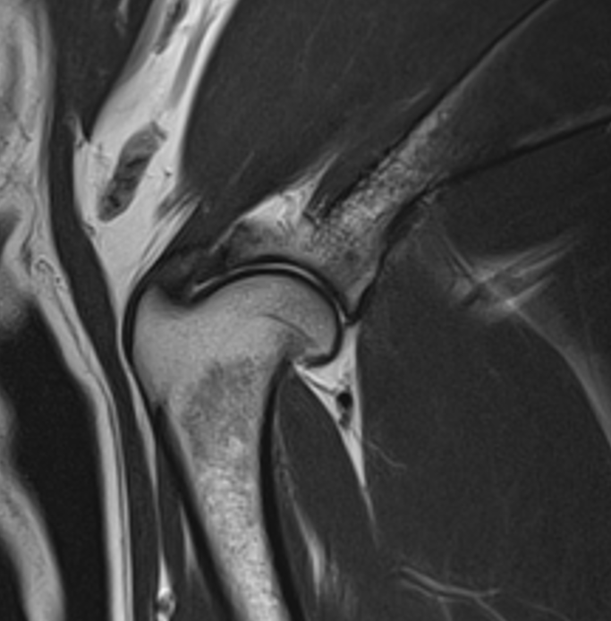
- One of the many benefits of an MRI is its ability to easily assess body parts and structures in three dimensions. The multi-plane imaging capability can lend this imaging modality well to animal athletes with complex injuries that may not be easily visible on a single image. This comprehensive and three-dimensional view of the injured area can aid veterinarians in making a better-informed decision and, ultimately, improved treatment and rehabilitation recommendations. For example, in the case of this 2-year-old Rottweiler with left thoracic limb lameness suffering from pain on extension of the shoulder. An MRI study was performed showing:
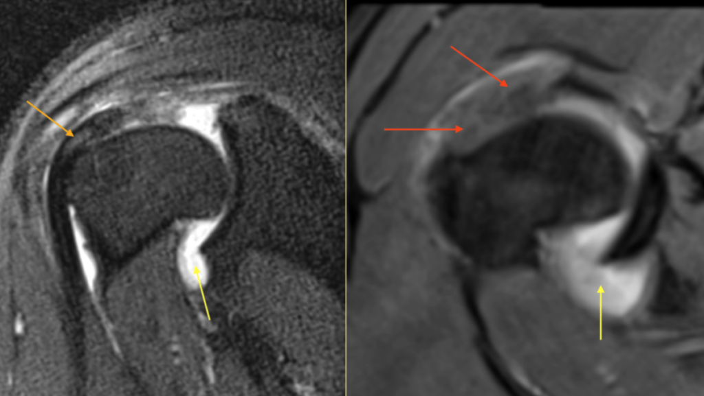
The red arrows point to a markedly swollen supraspinatus tendon, causing medial displacement of the biceps tendon (orange arrows). Notice that the bicep is mildly hyperintense. The yellow arrow points to joint effusion within the bicipital tendon sheath. Based on the MRI study a supraspinatus tendinopathy was suspected, causing secondary biceps tendinitis and joint effusion
Taking Care of Our Four-Legged Friends
The impact of animals on our daily lives cannot be overstated; from four-legged family members to vital parts of our working and sporting lives. Animals have been our close companions in many of our daily endeavors and for millennia they have served instrumental roles in our societies. This is a testament to the close bond between humans and their animal companions. Like their human counterparts, these animals can suffer musculoskeletal injuries that can impair mobility and function, ultimately impacting their quality of life. If injuries are unrecognized and untreated, they can have a long-lasting impact on musculoskeletal health. In many cases, these injuries are complex and can prove challenging to diagnose effectively without advanced diagnostic tools, like an MRI.
INTERESTED IN VISIONARY VETERINARY IMAGING?