Magnetic Resonance Imaging (MRI) provides invaluable information about soft tissues and the identification of bone fluid, as well as other aspects of bony remodeling or injury. In addition, rescan MRIs are beneficial and especially vital in those cases where the lesions cannot be assessed, or are difficult to assess, with other imaging modalities such as radiography and/or ultrasonography. Elisabeth van Veggel, DVM, Dipl ECVDI-LA, from Sporthorse Medical Diagnostic Centre (SMDC) in Heesch, The Netherlands, discusses the value of rescan MRIs in aiding lameness diagnosis for your horse.
MRI Follow-Up Scans and Their Value
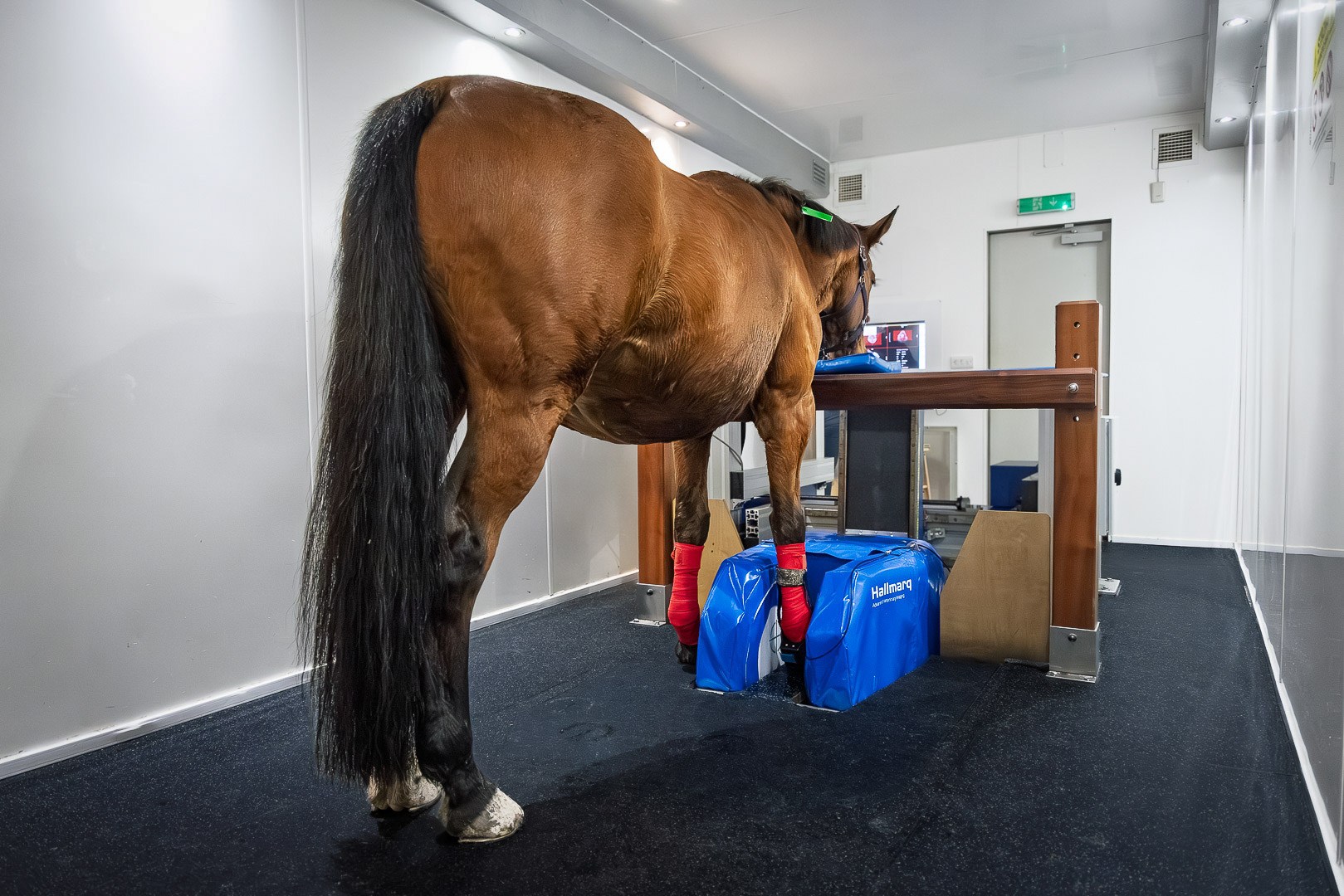
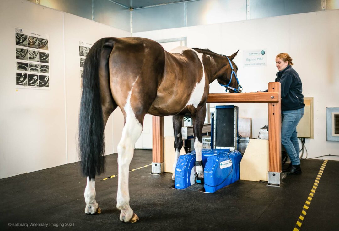
Standing Equine MRI can evaluate the limb from the foot to the carpus in the front limb and from the foot to the tarsus in the hind limb. For every lameness case, an accurate diagnosis is important to estimate prognosis and help guide and implement an appropriate treatment and rehabilitation plan. Furthermore, follow-up examinations, which may include rescan MRIs, have tremendous added value in monitoring lesions, guiding the rehabilitation and treatment plan, and further evaluating the prognosis (Werpy 2012).
Rescan MRIs are beneficial and are especially vital in those cases where the lesions cannot be assessed, or are difficult to assess with other imaging modalities such as radiography and/or ultrasonography.

MRI Where Other Modalities Fail
MRI has been used extensively in the foot to fetlock region for many years. It is also an excellent diagnostic aid for proximal metacarpal pain especially when other modalities, such as radiography and/or ultrasonography fail to identify the cause for lameness. Within the proximal metacarpal region, concurrent bony and soft tissue injuries are common (Murray et al 2020, van Veggel et al 2021). A previous study indicated that the severity or type of the lesions of the proximal metacarpal region does not influence the prognosis and that approximately 60% of horses returned to their previous work level (van Veggel et al 2021).
In the face of rescan MRIs, the evolution of the lesion is important. Certain lesions are expected to improve, some are expected to stay the same, and certain lesions could look worse on the follow-up MRI. Whatever the case, both the initial clinical examination and any follow-up clinical examinations, are important and should be taken in conjunction with the MRI findings.
The Value of MRI Rescans as Proven by Clinical Evidence
A recent study – published by SMDC – evaluated a group of horses with proximal metacarpal pain that returned to their previous level of work (van Veggel 2024). In this small group of horses, resolution of the third metacarpal bone fluid-like material (hyperintense STIR signal) is expected for horses returning to soundness (van Veggel 2024). This is an important finding and indicates that horses that are sound at the recheck clinical examination but still have bone fluid-like material in the proximal metacarpus at the recheck MRI may need more time to heal.
On the other hand, in less favorable situations, horses that are not improving clinically and have persistent bone fluid-like material in the proximal metacarpus may, unfortunately, be progressing toward chronic lameness. This injury (hyperintense STIR signal) cannot be assessed via radiography and ultrasonography thereby highlighting the importance of rescan MRIs.
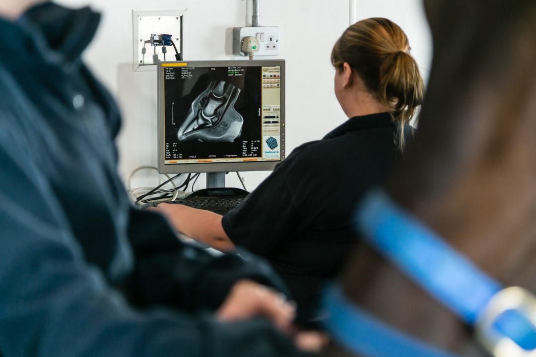
In addition, complete normalization of the soft tissue structures (the dorsal collagenous part of the proximal suspensory ligament), does not appear necessary for a return to soundness. Slightly more than half showed an improvement in the gradation of their injury but more than 1/3rd stayed the same. This area of soft tissue injury can be difficult to assess on ultrasound and in many cases the injury is more clearly and accurately seen on the MRI.

MRI Rescans and Timing
The time frame for any MRI recheck exam must be guided by several factors: the initial lesion, the time frame of the lameness, and the clinical progression. In addition, the owner’s financial considerations or, in some cases, the level of insurance cover, will also help determine the timeframe for an MRI rescan. A broad timeframe, often used in rescan MRIs, is approximately 90 days but the time frame is further finetuned on a case-by-case basis.
Conclusion
All in all, MRI plays an important role in the diagnosis of proximal metacarpal pain, and rescan MRIs will help estimate the prognosis and guide the treatment and rehabilitation plan.
INTERESTED IN VISIONARY VETERINARY IMAGING?


