Historically, a pre-purchase examination (PPE) for horses primarily consisted of a thorough clinical assessment. In addition to standard radiographs, Magnetic Resonance Imaging (MRI) is increasingly being utilized as part of the pre-purchase scenario. Guest author Maty Looijen, MSc MVetMed DipECVDI MRCVS, discusses the pros and cons of its use and the importance of decisions being made based on a clear understanding of the potential risks and benefits associated with imaging findings.
MRI and the Importance of Interpretation
It has become increasingly common to include radiographic evaluations as part of the PPE process. In addition to standard radiographs, discussions around the utility of ultrasonography and additional radiographs – such as those of the neck and back – have emerged. Notably, magnetic resonance imaging (MRI) is being utilized more frequently in these examinations. MRI has demonstrated its invaluable role in diagnosing lameness, aiding in prognosis, rehabilitation planning, and specialized treatment options. Nonetheless, questions arise regarding the interpretation of MRI findings in horses that appear clinically healthy.
Collaborative Decision Making for Best Results
Imaging during a pre-purchase examination is crucial for assessing potential risks associated with future performance or resale value. The interpretation of imaging results should be integrated with findings from the clinical evaluation and the horse’s known show record, if available. This process often involves collaboration among the examining veterinarian, the buyer, the seller or rider, and potentially veterinary specialists experienced in equine diagnostic imaging.
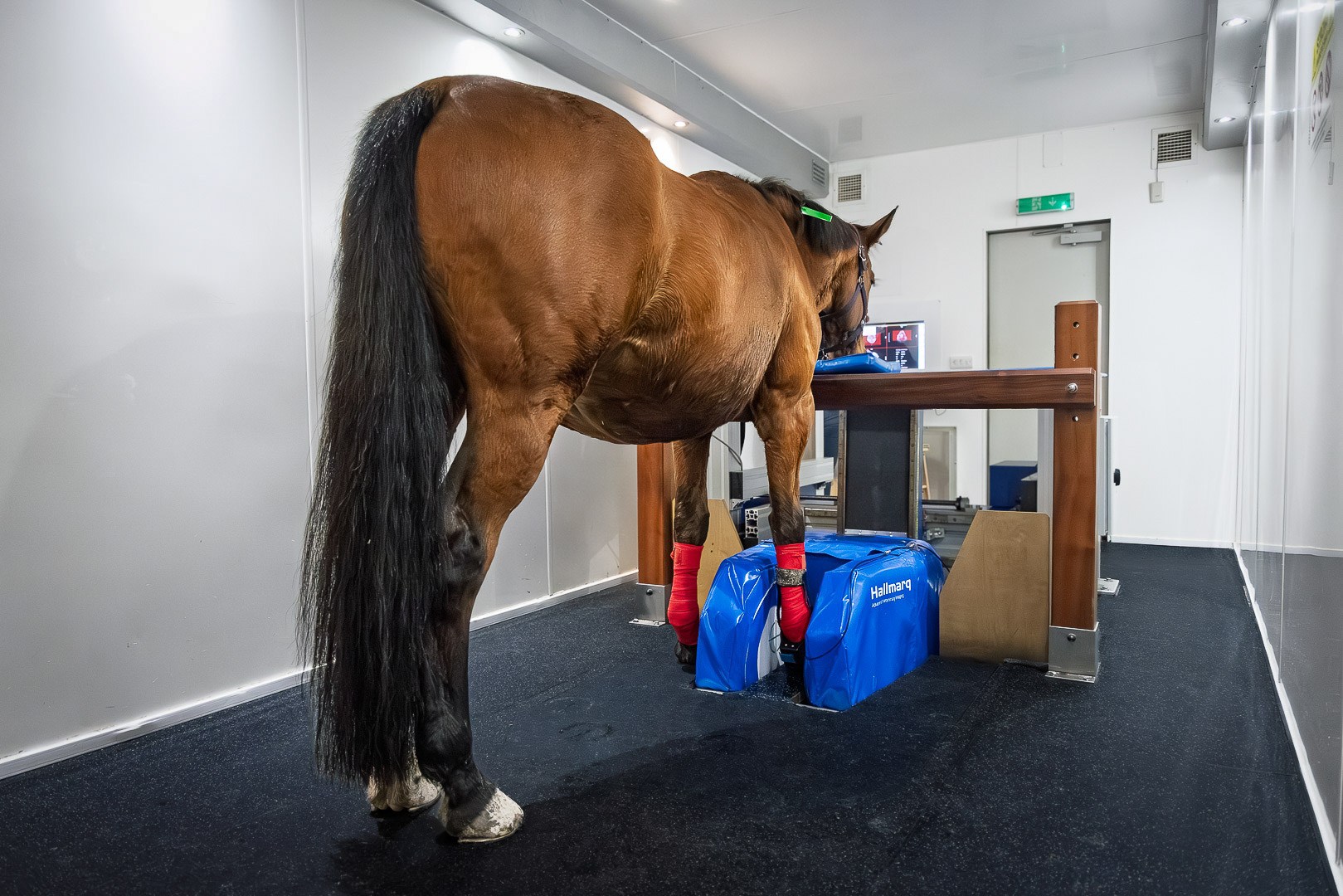
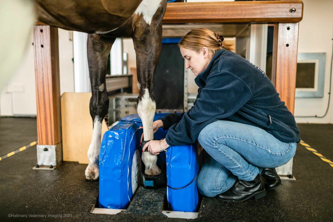
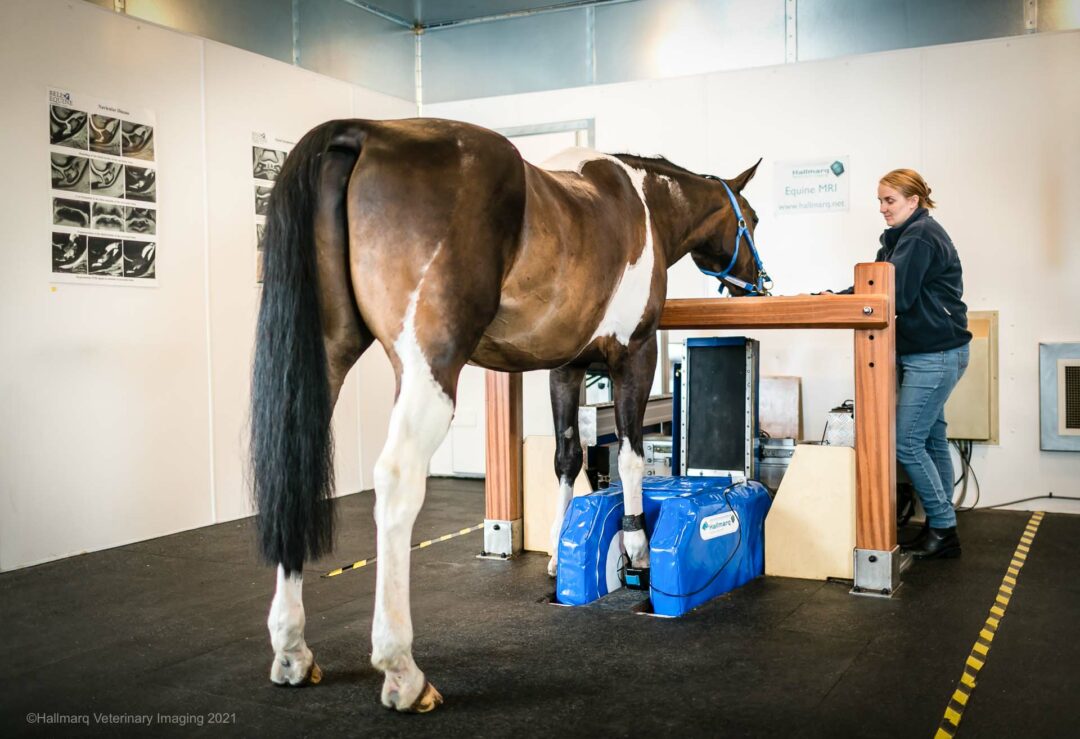
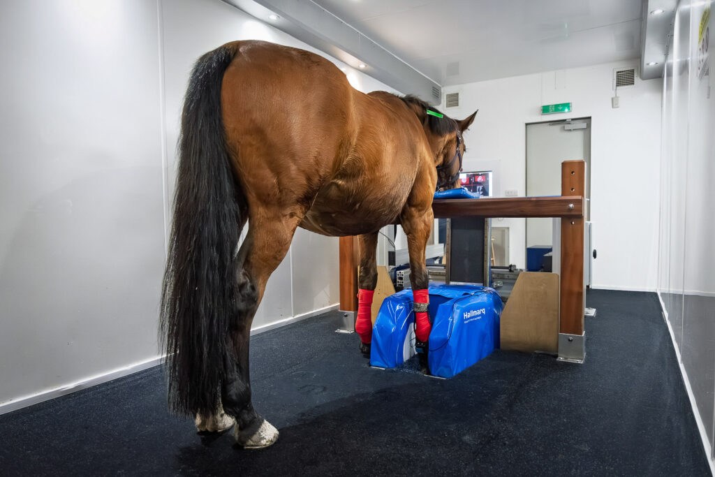
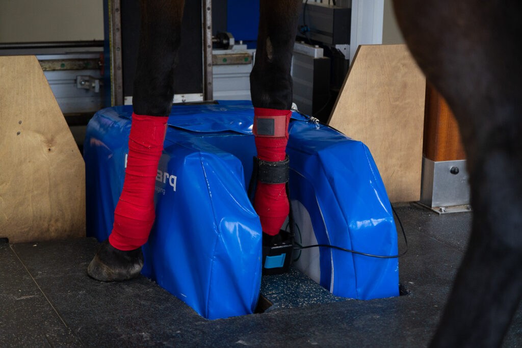
Standing MRI of the Foot for PPE
Radiographic examinations are the most common imaging performed and an integral part of the prepurchase examination. Radiographs are obtained to find performance-limiting or potential performance-limiting orthopedic problems. This however requires accurate diagnosis and interpretation of the images presented. When questionable findings arise, further diagnostic imaging investigation may be warranted. Standing MRI of the foot is particularly common in pre-purchase scenarios, as it can alleviate or confirm buyer concerns when radiographic changes fall outside expected norms based on breed, age, and discipline (Selberg, 2018).
MRI and Possible Misinterpretation
While there are clear benefits to utilizing MRI in pre-purchase examinations, it can also lead to complications—often referred to as opening a “can of worms” (Sherlock, 2015). The advantages and disadvantages of MRI must be thoroughly communicated to both the purchaser and vendor to facilitate informed decision-making. A significant challenge lies in the radiologist’s ability to interpret the clinical significance of imaging findings accurately. Radiologists may lack comprehensive knowledge about the aging and progression of specific lesions, necessitating extensive histopathological validation and follow-up studies on sound horses. There are lesions, such as moderate and severe DDFT tendinopathy (Garcia et al., 2013), which are clearly described however, the majority of low grade findings are not.
MRI and Individual Susceptibility
Interestingly, numerous high-level competition horses may exhibit imaging abnormalities without any signs of lameness. This suggests individual susceptibility to lameness resulting from these lesions. Moreover, radiologists often do not possess sufficient expertise to predict how subtle diseases will progress based on MRI findings. Misinterpretations stemming from lack of scientific evidence could expose radiologists to liability if a horse subsequently becomes lame.
Balancing MRI Results With Clinical Relevance
The ambiguity surrounding many imaging findings can lead to both underestimation and overestimation of their significance. Consequently, excessive reliance on MRI may result in horses with the potential for long and successful athletic careers being sidelined due to misinterpretations of clinical significance. The difference in between findings of a normal sound horse and a lame horse are sometimes only minimal.
In a study performed by Murray et al, they found differences in severity in between those with palmar foot pain and those without any clinical problems. However, still in the control group 27% of horses shows moderate changes (Murray et al., 2006). One proposed solution is for this is to report only the imaging findings without attempting to assess their clinical relevance. While this approach may mitigate issues related to interpreting clinical significance, it likely will not alleviate client concerns (Sherlock, 2015).
Improved Understanding for Best Results
A study performed in 2020 on sound horses revealed that 3T MRI findings of the navicular bone had a good agreement to histopathology. The question remains however what are the agreeements comparing to standing low field MRI, which is obviously more appropiate in a prepurchase setting, and what is the clinical significance of those findings (now and in the future). Sherlock emphasizes the need for further studies involving sound horses and additional follow-up research to enhance our ability to interpret MRI results effectively during pre-purchase examinations (Sherlock, 2015). Until our understanding improves significantly, it is advisable that MRI be reserved for special circumstances with well-informed clients and careful interpretations.
In Conclusion
In conclusion, while MRI represents a powerful tool in equine diagnostics—particularly for lameness—it is essential that its use in pre-purchase examinations is approached with caution. Comprehensive communication among all parties involved is critical to ensure that decisions are made based on a clear understanding of potential risks and benefits associated with imaging findings.
Images © Hallmarq Veterinary Imaging 2025
