Spinal MRI: A Highly Effective Diagnostic Tool
One of the most reliable ways to detect spinal disease in pets as a cause of weakness is through spinal MRI (Magnetic Resonance Imaging). This advanced diagnostic tool provides highly detailed spine images, allowing veterinarians to visualize the spinal cord, nerves, surrounding spinal bones, and their muscles. With this precise 3D imaging, veterinarians can better understand the extent of most diseases and develop the most appropriate treatment plan. Magnetic resonance imaging for pets is beneficial because it provides a non-invasive method to assess complex spinal structures that are not easily detected through traditional X-rays or CT scans.
Conditions Typically Discovered with Spinal MRI
A spinal MRI can reveal a variety of conditions, such as:
- Intervertebral disc disease (IVDD)
- Infections and inflammation
- Spinal “strokes”
- Spinal tumors
- Spinal cord compression
This makes MRI an invaluable tool in diagnosing a wide range of spinal issues in pets at an early stage.
The Importance of Early Spinal Tumor Detection in Pets
These tumors can cause a range of symptoms, from subtle changes in mobility to severe pain and paralysis, which, if left untreated, can drastically reduce a pet’s quality of life. By identifying spinal tumors early, veterinarians can intervene more effectively, potentially improving prognosis, reducing pain, and preserving mobility. Early detection allows for timely treatment that can prevent further damage to the spinal cord and surrounding structures, leading to a more successful outcome for the pet.
Hallmarq: Leading Provider of Veterinary Imaging Solutions
Hallmarq is a trusted leader in advanced veterinary imaging, offering innovative MRI systems specifically designed for animals and their unique anatomy. Their cutting-edge technology improves pets’ health and well-being by enabling early detection and accurate diagnosis. Hallmarq’s systems are tailored to meet animals’ unique anatomical and diagnostic needs, ensuring the best care possible.
In this blog, we will further explore how spinal MRIs and early detection can enhance the quality of life for your dog or cat.
Why Spinal MRI Is Crucial for Tumor Detection
Understanding Spinal Tumors in Pets
Spinal tumors in pets are abnormal growths that can develop within or around the spine, affecting the spinal cord, nerves, and surrounding tissues such as bones and muscles. These tumors can be benign or malignant, and their severity depends on their specific type, size, location, and how quickly they grow.
Spinal tumors can cause a range of symptoms including:
- Mild discomfort
- Weakness
- Severe pain
- Paralysis
- Loss of coordination
As the tumor grows, it may compress the spinal cord, leading to neurological deficits that impact an animal’s ability to move, stand, or walk. Spinal tumors can significantly reduce a pet’s mobility and overall quality of life without timely intervention, making early detection crucial. Veterinary diagnostic imaging plays a pivotal role in effectively identifying these conditions.
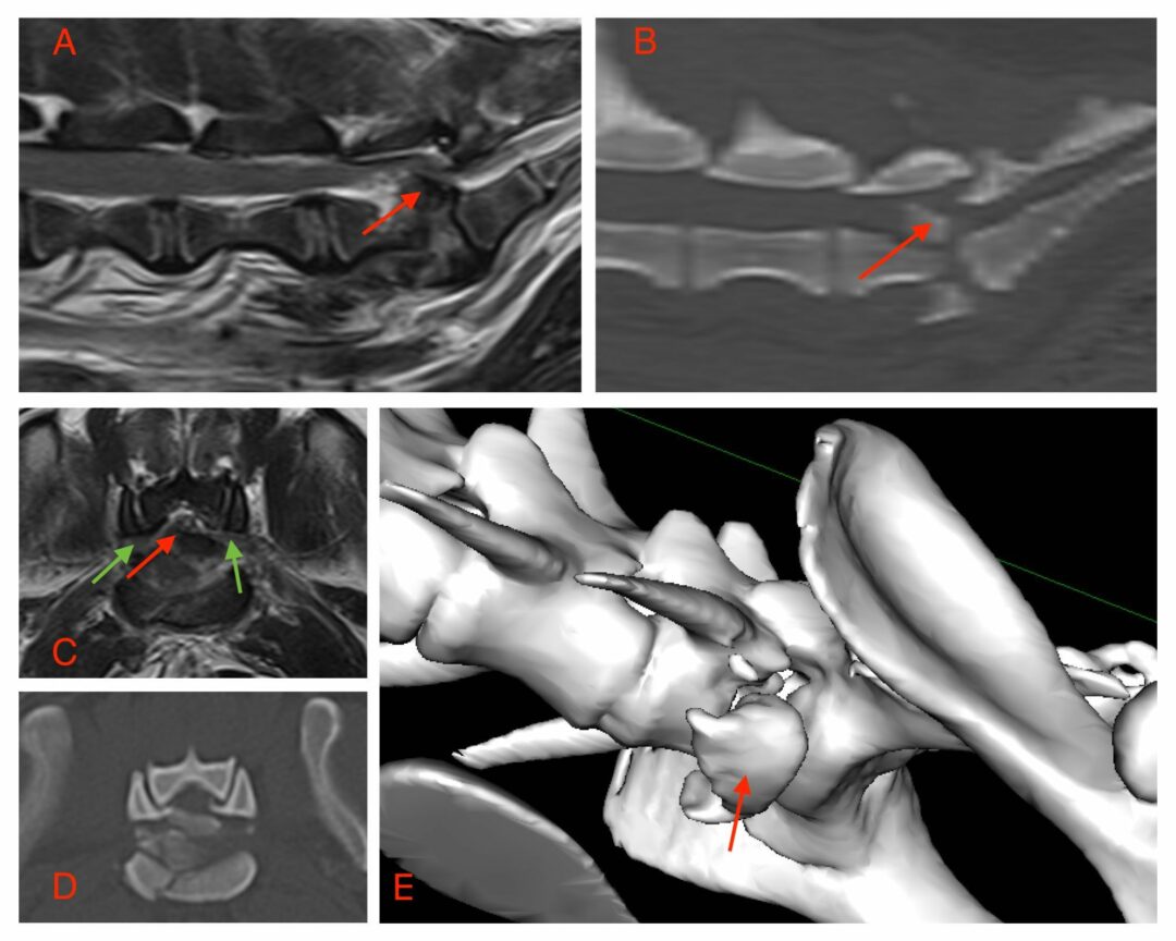
The image on the right shows a cross-sectional MRI of a vertebra clearly demonstrating a large irregular lesion associated with the vertebral body (arrow) and compressing the spinal cord. The lesion was confirmed to be a bone tumour called chondrosarcoma.

MRI vs. Other Diagnostic Methods
MRI (Magnetic Resonance Imaging) is the most precise and effective tool when diagnosing spinal tumors and related conditions in pets. While traditional methods such as X-rays and CT (Computed Tomography) scans are valuable for identifying bone abnormalities, they often fall short in assessing soft tissues like the spinal cord, nerves, and muscles.
X-rays primarily capture images of bones, making them helpful in detecting fractures or other skeletal issues. Still, they cannot provide detailed images to assess soft tissue structures or detect small tumors. CT scans offer more detailed images than X-rays and can show some soft tissue contrast in 3D. However, they still lack the resolution to fully visualize the intricate details of the spinal cord and surrounding tissues.
In contrast, MRI provides superior soft tissue imaging, allowing veterinarians to detect even small spinal tumors and assess their impact on nearby structures more precisely. This makes MRI the gold standard for diagnosing spinal issues that involve soft tissues, including tumors, herniated discs, and inflammation.
The Benefits of Early Detection with MRI
One of the most essential benefits of spinal MRI is its ability to detect tumors early, even before symptoms become severe. Early detection significantly improves treatment outcomes, allowing veterinarians to intervene sooner, potentially slowing the tumor’s progression and minimizing damage to the spinal cord. This can help preserve an animal’s mobility, reduce pain, and improve their overall quality of life.
In cases where surgery or other treatments are needed, early diagnosis through MRI gives veterinarians the information they need to plan more effective and targeted treatments. MRI can also extend a pet’s life expectancy by catching spinal tumors in the early stages. Timely treatment can prevent further deterioration and allow pets to enjoy a longer, healthier life with fewer complications.
Signs Your Pet Might Need a Spinal MRI
Spinal disease in pets can be difficult to detect early on, but specific symptoms may indicate a problem that requires further investigation. If you notice any of the following signs, it could suggest that your pet has a spinal disease, which in some cases could be a serious condition like a spinal tumor:
- Limping or Lameness: Favoring one leg or struggling with coordination may indicate nerve or spinal compression.
- Difficulty Walking or Stumbling: Unsteady movements or dragging back legs could point to spinal problems.
- Sensitivity Along the Spine: Pain or sensitivity when touched along the spine may signal an underlying issue.
- Visible Pain: Yelping, whimpering, or flinching when moving are common indicators of spinal discomfort.
Behavioral Changes as Early Indicators
- Reduced Activity: A once active pet may become lethargic.
- Reluctance to Jump or Climb Stairs: Pain may cause hesitation in usual activities.
- Loss of Appetite or Mood Changes: Irritability or restlessness can be signs of spinal discomfort.
When to Consult Your Veterinarian
If you notice any of these signs, consult your vet. Your veterinarian may recommend a spinal MRI to identify the root cause of discomfort. Early detection can ensure your pet gets the proper treatment and care.
Conclusion
Spinal MRI has revolutionized veterinary medicine by offering unparalleled diagnostic imaging capabilities for various conditions. Advanced tools like MRI help detect issues in pets with exceptional precision, improving outcomes. Always consult your veterinarian to determine the best approach for your pet’s unique needs.
In our next blog, we’ll provide a step-by-step guide to spinal MRI for cats and dogs.