


When traditional lameness diagnostics like nerve blocks, radiographs and ultrasound don’t provide clear answers, advanced imaging can make all the difference. Standing Equine MRI offers unparalleled insight into both bone and soft tissue without the risks of general anesthesia. For equine vets, knowing when and why to refer for MRI can transform outcomes, improve client confidence, and potentially prevent equine athletes from career-ending injuries.
Why Standing Equine MRI is Essential For Lameness Diagnosis
You’ve done the lameness work-up: trotted, lunged, seen under saddle, performed nerve blocks, taken radiographs, and maybe even scanned with ultrasound. And still… limited answers.
We understand the frustration! You’ve done everything right but the horse is still lame, the owner is growing impatient and the pressure is mounting. You want to help but need further clarity on the diagnosis.
So what’s the next step?
Advanced Imaging With Standing Equine MRI
Only MRI can detect both bone and soft tissue abnormalities that radiography and ultrasound may miss, helping you make confident decisions and deliver better outcomes for your patients.
Hallmarq MRI is one of the best decisions we’ve ever made. It’s opened up avenues for diagnosis and therapies we never thought possible.”
Rick Mitchell, DVM, MRCVS, DACVSMR, Cert. ISELP, Fairfield Equine, CT
Let’s look and why, when and how to refer for Standing Equine MRI.
Why Refer for Standing Equine MRI?
Because advanced imaging with Standing Equine MRI means you can be right 90% of the time!*
MRI is the only modality that visualizes both soft tissue and bone in exquisite detail and without any of the risks associated with general anesthesia. It’s ideal for:
- Multifactorial lameness where X-ray and ultrasound fall short
- Diagnosing conditions like navicular syndrome, collateral desmitis, and bone marrow edema
- Monitoring healing and assessing readiness for return to work
MRI reveals inflammation, bone bruising, and subtle lesions that are invisible on other modalities. If a horse responds to a palmar digital or coffin joint block, it’s a strong candidate for MRI of the foot.
For abaxial sesamoid block responses, scanning the pastern or fetlock isn’t a problem. New iNAV motion correction software means imaging higher up the limb is now far easier, removing motion artifact to deliver transformational results for a definitive diagnosis.
With MRI, you can detect early subchondral bone and cartilage lesions, allowing for rest and training adjustments before catastrophic injury occurs. For the athletic competition horse, continued performance training to keep in peak condition inevitably results in occasional injury. Early detection of soft tissue problems and bone changes in response to stress enables training adjustments before an injury progresses to a catastrophe.
When to Refer for Standing Equine MRI
When you need more information, that’s the time to refer:
- Nerve blocks localize the pain, but imaging is inconclusive
- Ultrasound access is limited, or anatomy is complex
- You want to avoid general anesthesia
- You need to track healing or plan a return to competition
How to Refer for Standing Equine MRI
The referral process is straightforward:
- Contact the referral clinic with case details
- The clinic handles scheduling and client communication
- You receive a radiological report and interpretation
- A client-friendly summary to support your treatment discussion
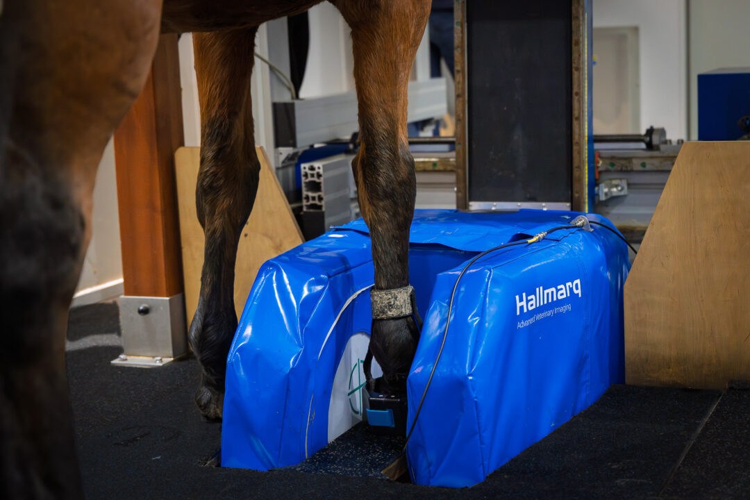
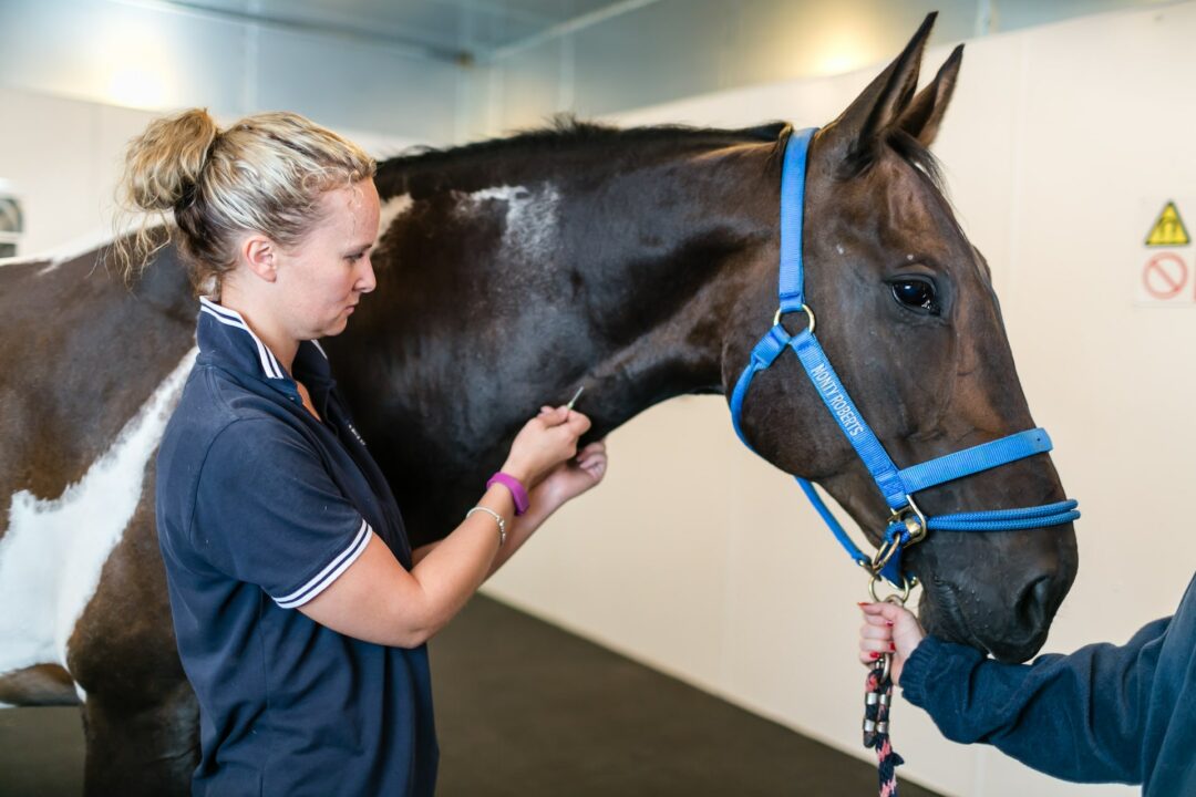
What to Expect During a standing MRI Procedure
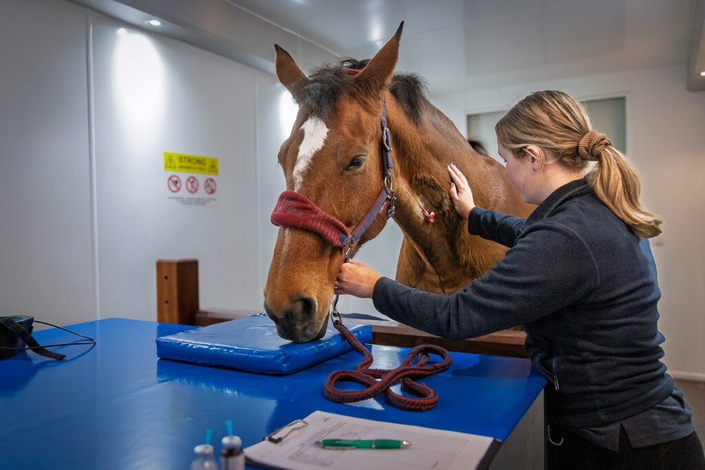

- A safe, standing, sedated procedure without general anesthesia
- A scan produces somewhere between 300–500 images
- Enhanced motion correction software ensures clarity
- Full evaluation of soft tissue and bone lesions
Conclusion
You’re not alone in the diagnostic journey. Standing Equine MRI is uniquely placed to support your clinical investigation, uncover hidden pathology, and get horses back to work. When the usual tools aren’t enough, MRI is your next step forward.
Standing Equine MRI: The Difference Between Guessing and Knowing!
*Reference: Koch DW, Barrett MF, Jackman BR, MacDonald D, Goodrich LR. (2020) Comparison of lameness outcomes in horses with acute or chronic digital lameness that underwent magnetic resonance imaging. N Z Vet J. 68(5):283-288.