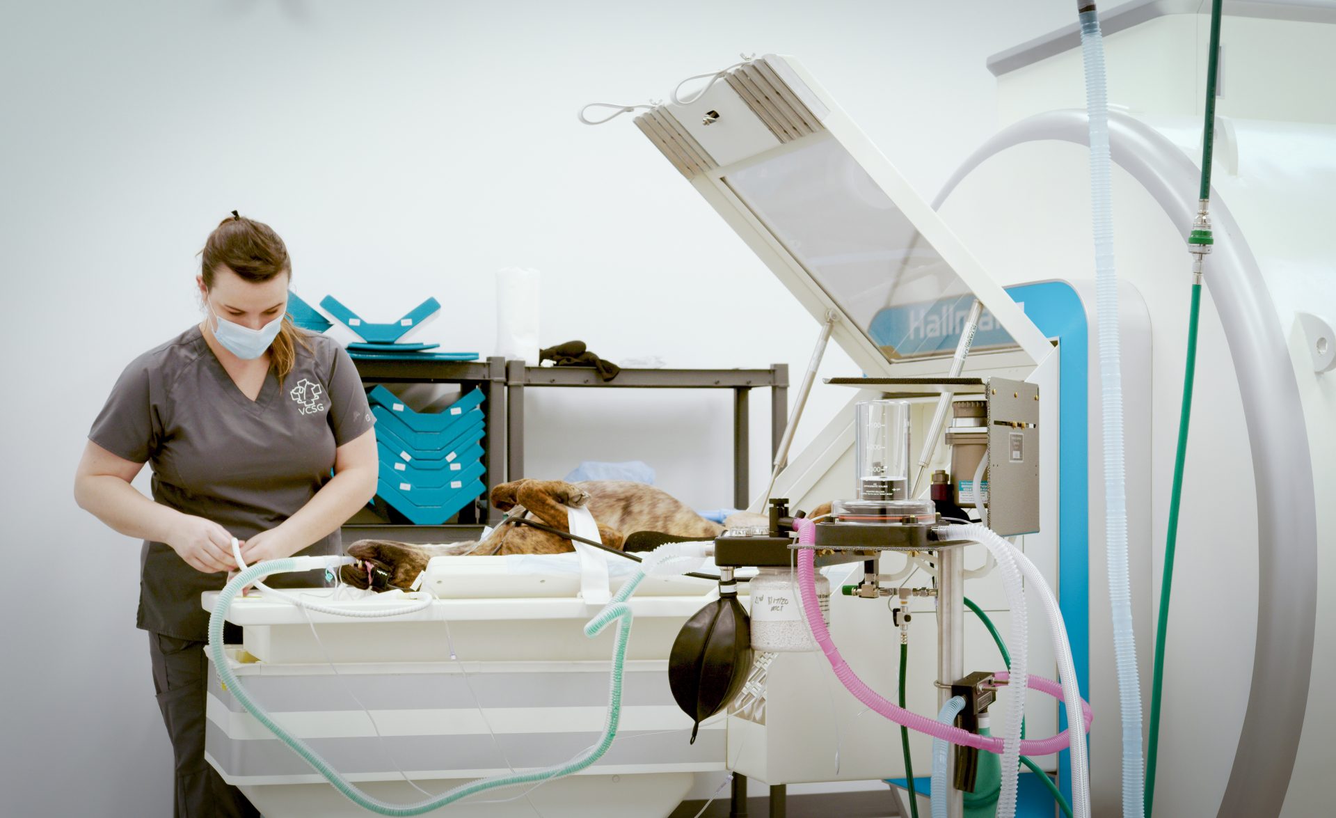
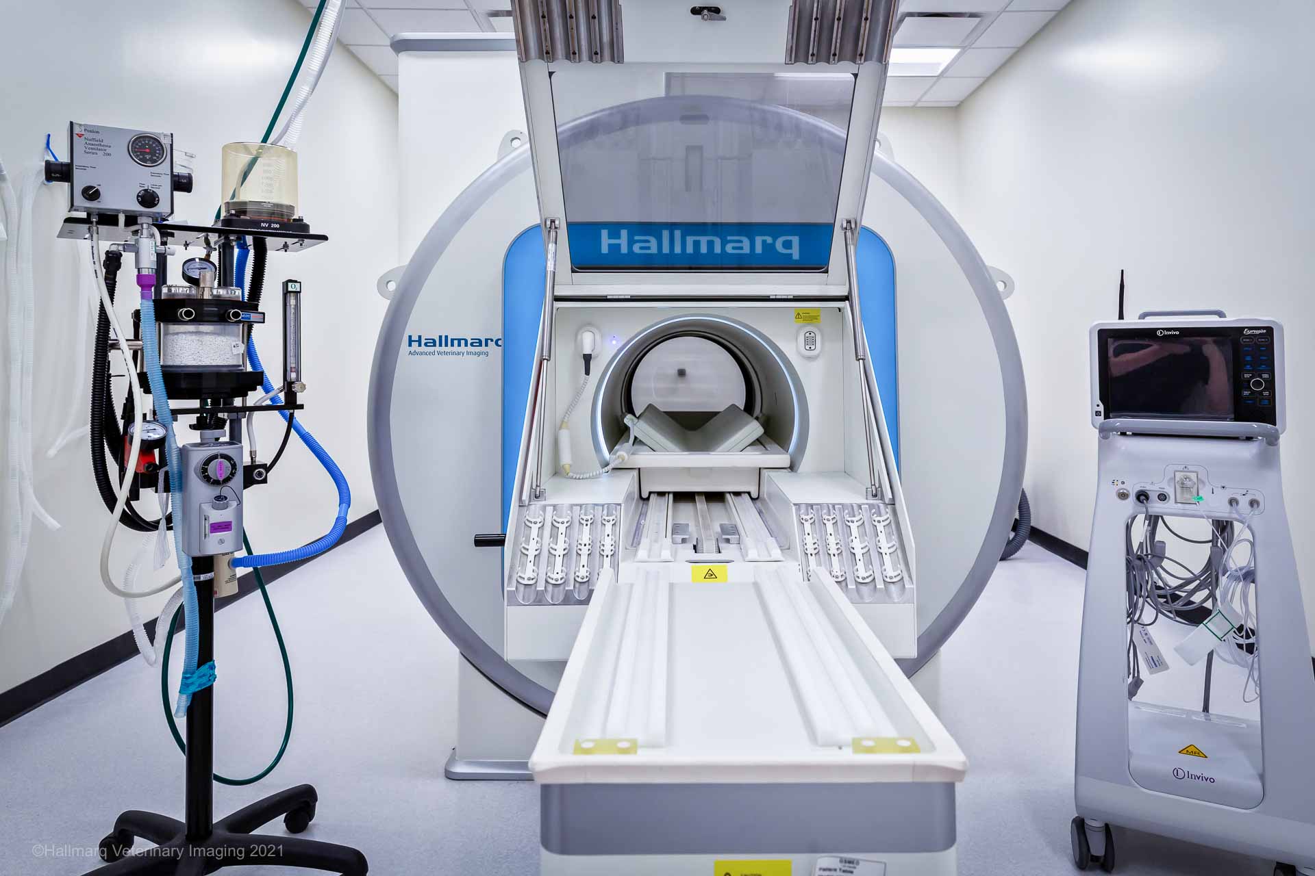
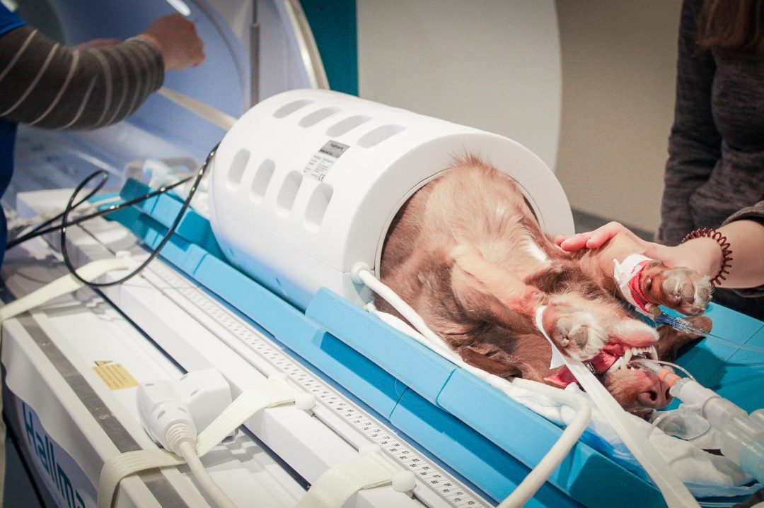
There are more applications for a small animal MRI system in a first opinion veterinary practice than most practitioners might realise. Offering the option of an on-site MRI service to a pet owner can help them make a more informed decision regarding their pet’s diagnosis and treatment.
Although MRI scans can be a significant expense for owners, insurance may cover most of the cost and the results help veterinarians pinpoint the root causes of these common symptoms you see patients presenting with every day. Here are the most common clinical use cases for your small animal MRI system.
Seizures
Seizures are often caused by epilepsy, a chemical disease that doesn’t show up on MRI scans. The outlook for epilepsy is good, but confirming the diagnosis requires eliminating other causes like brain tumor, stroke or inflammation which can be detected. You can treat seizures with epilepsy drugs without performing an MRI scan, but if they don’t work, it could point to a structural disease or inflammation that will require an MRI to diagnose and provide a more accurate treatment plan.
MRI scans show structural changes in the brain, whereas CT scans may appear normal and miss subtle brain abnormalities in cases with strokes, inflammation or encephalitis and tumors. If there’s nothing identifiable on an MRI image, then you can be more confident in your epilepsy diagnosis. Using your MRI system for these seizure cases helps increase confidence in diagnosis and treatment.
Balance Problems
MRI can be used to confirm the cause of the sudden onset of a head tilt or symptoms of ataxia, like a ‘drunken’ gait. These vestibular signs are among the primary symptoms of severe inner ear disease.
However, vestibular disease can also be caused by brain problems, which could be significantly more severe and require more expedient diagnosis and treatment. MRI is the only imaging test to help veterinarians give an accurate answer for the cause of severe balance issues.
Ear Diseases
Ear diseases that are long-standing and not responsive to treatment are another common use case for MRI systems. Progressive ear disease may result in balance problems but could also just be associated with regional pain and or facial paresis (indicated by a facial droop).
Pet owners may opt to treat the symptoms of ear disease with antibiotics first. But if antibiotics don’t work, refer for an MRI scan to either confirm ear disease or determine another cause for the symptoms that may require surgery.
CT and radiographs can sometimes pick up ear disease, but they can also miss it in its early stages and can’t demonstrate the extent to which it may have entered the brain. MRI is extremely sensitive for this purpose and can determine the extent of disease within the ear cavities.
Spinal Pain
The two most common areas for spinal pain are in the neck and lower back, the latter being especially common in large dogs. You need to determine if the pain is originating in the patient’s hips or spine and whether there is nerve involvement. Spinal pain is frequently caused by a slipped disc, but there are several other potential causes that MRI can help rule out.
If your patient doesn’t respond to standard pain relief, then you’ll have to do a more extensive diagnostic investigation to redirect your treatment as soon as possible. CT can sometimes pick up disc problems, but usually misses its effect on nerves and spinal cord.
Occasionally the condition is within the spinal cord itself. Syringomyelia, for example, is a frequent cause of neck issues in toy breed dogs, especially Cavalier King Charles. Syringomyelia is often combined with Chiari-like syndrome, a congenital malformation of the skull.
These sources of head, neck and back pain can’t be picked up by CT. Most pet owners will opt for an X-ray first as the most cost-effective imaging technique, but if that is considered to be normal, then an MRI scan should be your next step. You can perform a CT next instead, but if that too is normal, you will still have to run an MRI scan to determine the cause of pain.
Acute Paralysis
In many cases, paralysis occurs suddenly and can result in partial or total loss of limb control or movement. A quick diagnosis is crucial to determine whether surgery could give the patient a chance for recovery. CT scans may appear normal, resulting in the need for an MRI to pinpoint the cause.
Owners often perceive paralysis as incompatible with good quality of life for their pets and may consider euthanasia. Since many will make a life-or-death decision for their pet, paralysis requires a quick diagnosis to determine whether it can be reversed or surgically corrected.
Behavioral Changes
Slow deterioration in behavior could indicate a serious brain abnormality or simply age-related changes. It’s important to understand age-related change versus other causes such as a brain tumor or encephalitis. When a pet presents with behavior changes, the most important diagnostic step to help the veterinarian achieve diagnosis is to consider an MRI.
Treatment will be more specifically guided by the results of an MRI study. Multiple brain diseases can cause the same signs, resulting in treating symptoms instead of uncovering the causes. These diseases could continue to progress but age-related causes could simply be addressed with a diet change! Your MRI system will support a more definitive diagnosis of the cause of behavioral changes, enabling you to pursue more targeted, and therefore successful, treatment options.


