Foot Penetrating Injury
In this case study, kindly provided by Hästklinik Florian Lackner, we take an in-depth look at a horse presented for an acute penetrating foot trauma from a rusty nail. Combination scanning using standing equine MRI and Vision CT was used to help reach a diagnosis and proved the usefulness of multi-modality imaging.
MRI Findings
The horse underwent a Standing Equine MRI to assess damage to the vital structures (such as the navicular bursa and deep digital flexor tendon). However, suspectable metal fragments from the rusty nail resulted in a large susceptibility artifact, obscuring the region of interest (Figure 1).

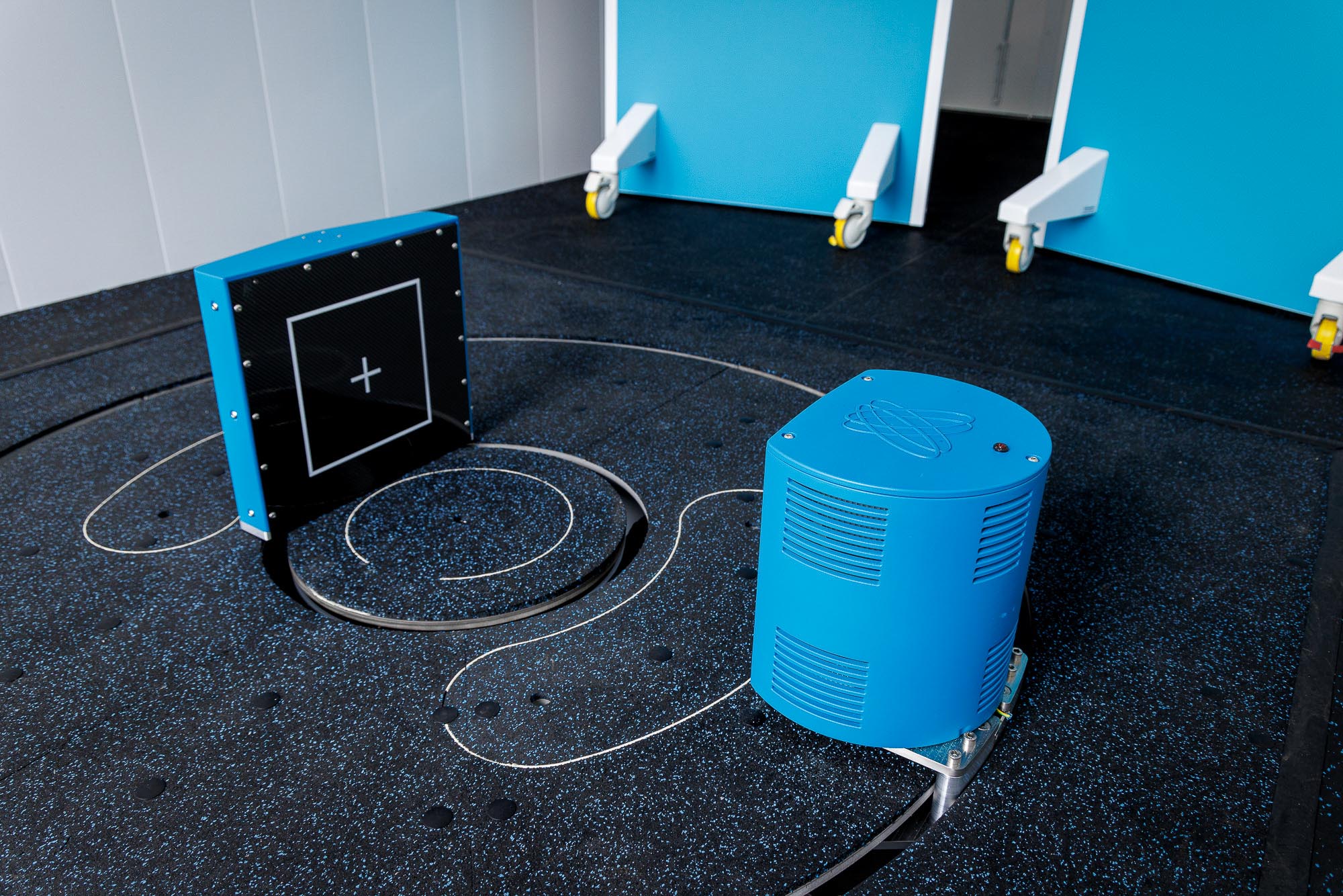
CT Findings
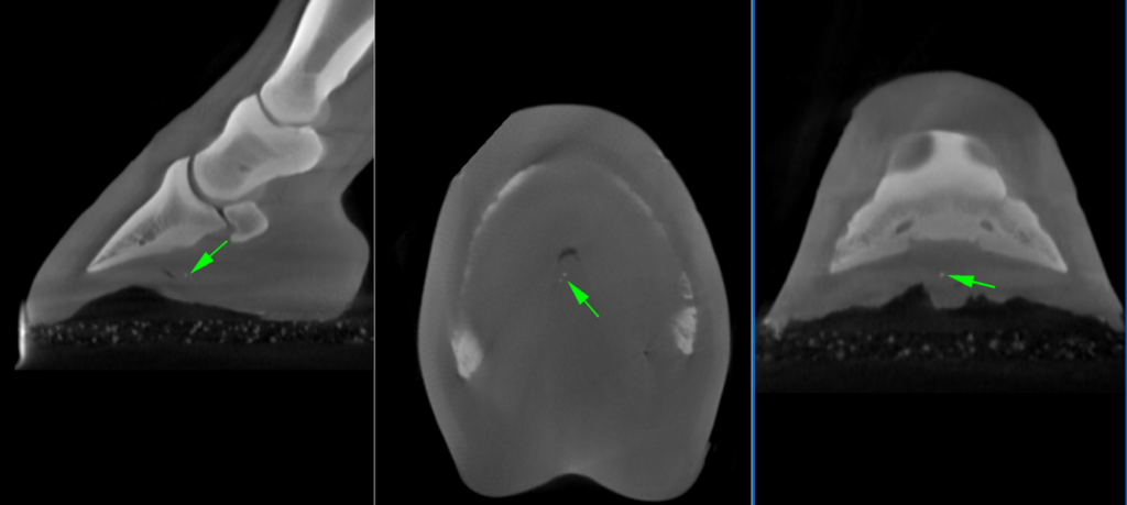
Utilizing Hallmarq’s 3D Vision CT imaging, the penetrating tract was successfully visualized (Figure 2) and revealed multiple metal fragments within the frog (Figure 3). The nail tract was relatively short and had a dorsal direction, with no involvement of vital structures detected.

Conclusion
MRI is a well-established diagnostic tool for foot penetrating injuries due to the three-dimensional foot evaluation. It provided the physiological data needed. However, in this instance, the presence of metal fragments limited the diagnostic efficacy of MRI. CT imaging – which provided additional structural data – proved instrumental in visualizing the tract and assessing the integrity of crucial internal foot structures.
With thanks to Louise Ovesen DVM, Hästklinik Florian L Ab, Sweden, for sharing this case with us.