When your pet suddenly starts limping or showing signs of lameness, it can be cause for great concern. While some cases may be resolved with rest or a trial of anti-inflammatory therapy, others require advanced diagnostic tools to uncover the underlying issue. One such tool, magnetic resonance imaging (MRI), is becoming increasingly valuable in veterinary medicine. Known for its precision and ability to provide detailed images of soft tissues, joints, and bones, MRI offers unique advantages in diagnosing lameness in pets
Understanding lameness in pets
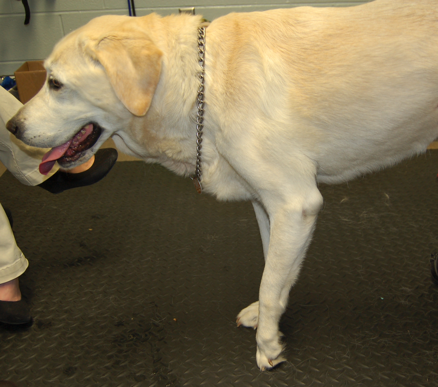
Lameness refers to an abnormal manner of walking or difficulty in moving due to pain, injury, or weakness in a limb. It can stem from various causes, such as joint, bone or neurological diseases. Diagnosing lameness can be challenging, especially when symptoms are subtle, or when standard diagnostic methods like radiographs fail to provide definitive answers, which is often the case with neurological diseases.
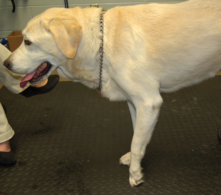
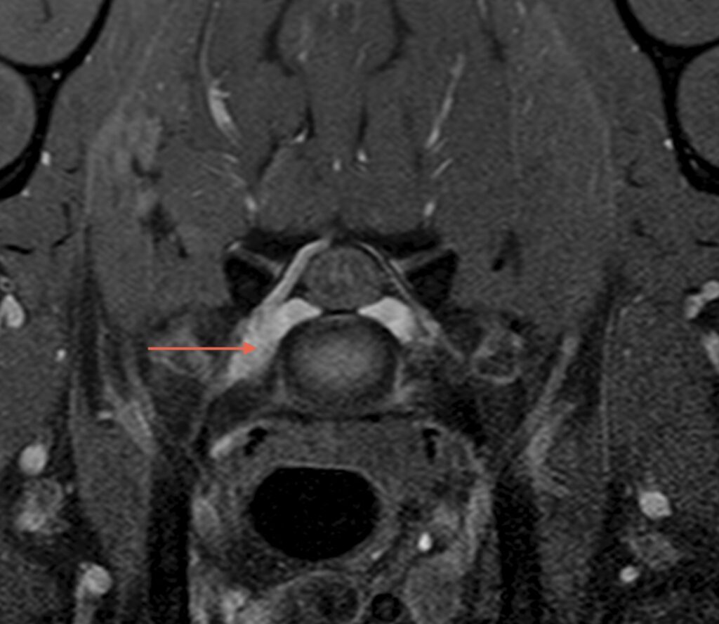
Unlike traditional imaging methods, MRI can provide cross-sectional images of the body, allowing veterinarians to examine soft tissues, cartilage, tendons, ligaments, nerves, and the spinal cord with unmatched clarity.
Benefits of MRI in diagnosing lameness in pets
- Detailed imaging of soft tissues: Lameness often arises from injuries or inflammation in soft tissues such as tendons and ligaments. Unlike radiographs, which primarily evaluate bone structures, MRI provides detailed images of these soft tissues. This is particularly helpful in diagnosing conditions like cruciate ligament disease, tendon injuries, or meniscal damage in joints.
- Assessment of Neurological Causes
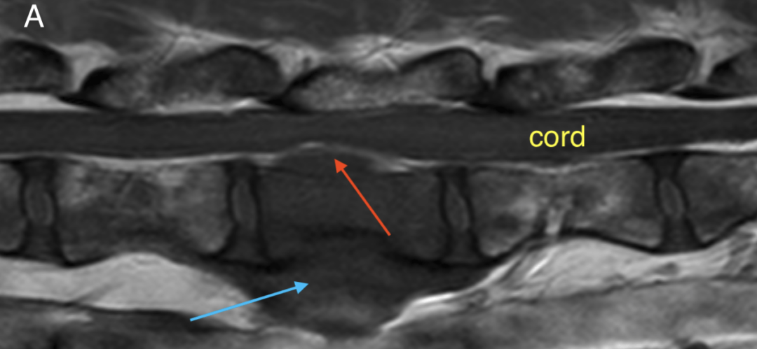
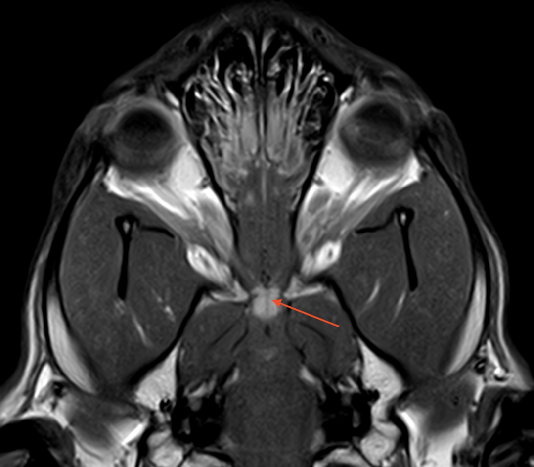
Some cases of lameness are linked to neurological issues, such as intervertebral disc disease (IVDD), nerve root compression sometimes called foraminal stenosis and nerve sheath tumours or trauma. MRI excels at visualizing the spinal cord, nerves, and surrounding structures, helping veterinarians pinpoint problems that may not be visible through other imaging techniques.
- Early Detection of Subtle Issues
MRI can detect early and subtle changes in tissues, such as inflammation, micro-tears, or early signs of joint degeneration. This early detection is critical for formulating effective treatment plans and preventing the condition from worsening.
- Non-Invasive Diagnosis
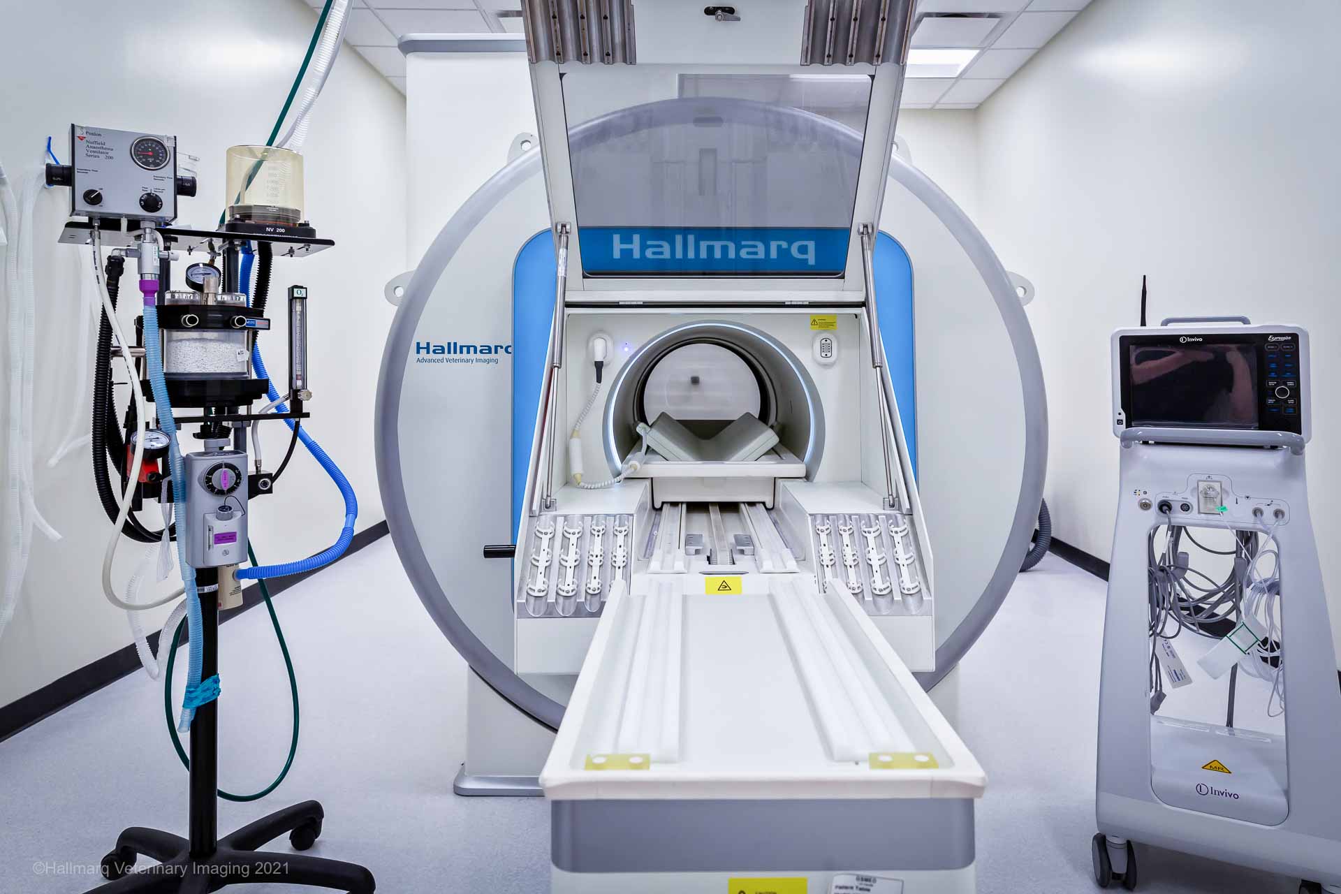
One of the biggest advantages of MRI is its non-invasive nature. It eliminates the need for exploratory surgeries, reducing risks and recovery time for your pet. Sedation or general anesthesia is typically required to keep the pet still during the procedure, but the process itself is painless.
Common applications of MRI in lameness diagnosis in pets
Veterinarians may recommend MRI for various conditions, including:
- Osteoarthritis: To assess joint cartilage and detect degenerative changes.
- Soft Tissue Injuries: For identifying tears or inflammation in tendons and ligaments.
- Vertebral, Bone and Joint Infections: To evaluate the extent of infection or abscesses.
- Tumors or Masses: To differentiate between benign and malignant growths that may affect mobility.
Conclusion
MRI is invaluable in diagnosing lameness in pets, which is not resolving with rest and anti-inflammatory medications, has not had a cause determined on radiographs or is neurological. By providing detailed, high-resolution images it enables veterinarians to identify the root cause of lameness and develop targeted treatment plans. While it may be more expensive than traditional imaging methods, its accuracy and ability to uncover a diagnosis often make it a worthwhile investment in your pet’s health. If your pet is struggling with lameness and standard diagnostics haven’t provided answers, consult your veterinarian about whether MRI could be the right option.
