


MRI has become a commonly used diagnostic imaging tool in diagnosing pathology of the distal limb of horses. With low-field MRI, patient motion has previously posed limitations on image quality when performing standing MRI in the proximal limb. For that reason, it has typically been less utilised in the equine field. But that’s no longer the case!
With increasingly experienced staff, and the shared knowledge of how to perform scans of the proximal suspensory, MRI should be considered a valuable tool in lameness cases of the proximal limb. It delivers a more complete picture of the injuries for a more accurate diagnosis. When paired with new advances in motion correction software – like Hallmarq’s iNAV – imaging of the proximal limb is now much improved. These new innovations enable us to make the best treatment and rehabilitation plan for the individual case.
Guest author and ECVDI resident – Rebecca Finne – currently works at Malaren Hastklinik, Sweden and writes of how best to utilise low-field MRI for bone injuries of the proximal limb in our equine patients.
MRI for bone injuries
MRI is known for its superior soft tissue details, but it also provides important information about bone pathology. Using different MRI sequences, you can evaluate and differentiate normal bone components like cortical bone, trabecular bone, and bone marrow. Adaptation to load, injuries, and pathological processes can cause changes in this normal bone composition and, depending on the process, cause either increased or decreased bone density, fluid replacing the fatty tissues or new bone formations (osteophytes, enthesophytes, periosteal, or endosteal new bone).
Looking at all these changes in the bone composition, you can evaluate the sclerosis, demineralisation, resorption, or fluid signal in the bone (“bone edema”) (Murray, 2010). MRI is currently the only imaging modality used in living horses providing visualisation of fluid signal in the bone (“bone oedema”), which is a particularly important part for evaluating bone injuries. (Murray, 2010).
However, limitations in bone evaluations with MRI also exist. It is not particularly sensitive to small pathologies, like fragments or osteophytes. This is linked to the slice thickness and contrast of the MRI. Combination with radiographs or CT is therefore helpful.
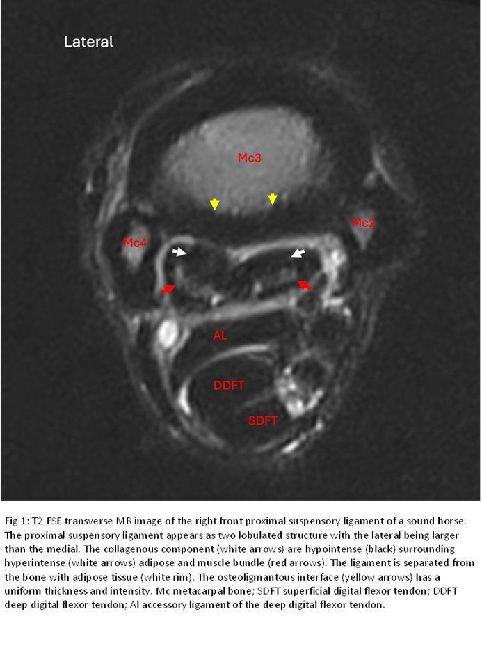
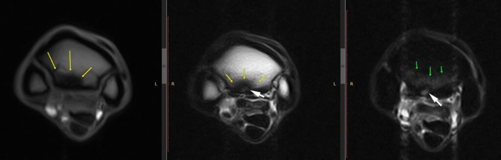
Proximal suspensory disease, looking further than just the ligament
Proximal suspensory disease is a complex pathological process in horses that involves more than just the ligament itself. The bone component can, in some cases, be more clinically significant than the ligament injury. At the suspensory ligament enthesis – the complex attachment of the ligament to the bone surface – the bone can have sclerosis, fluid signal (“bone oedema”), resorption or avulsion fragments (image 1), and fissures of the proximal metacarpal bone can also be identified on MRI in this region (Murrey et al. 2020, Labens et al. 2020).
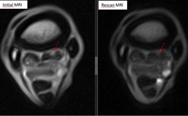
Combinations of suspensory injury alone or with a bone component may affect the rehabilitation programme for the horse (Ratcliffe et al. 2023). Following the fluid signal in the bone for lesions on repeat MRI scans can help determine the healing progress and optimize the timing of return to work (Van Veggel et al. 2024).
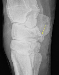
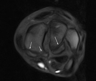
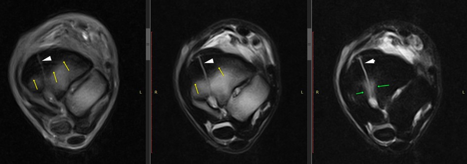
Carpal or tarsal bone fractures
Fractures of the carpal (image 2 and 3) or tarsal bones (image 4) are not always straightforward to diagnose with radiographs alone. MRI may safely – in a standing patient – provide an accurate diagnosis, precise fracture configuration, as well as assessing potential soft tissue injuries. This can be used for surgical planning of the fracture repair, or if conservative treatment is opted for, MRI is very useful for monitoring the fracture healing (Zimmermann et al. 2023).
MRI can further assess the position of these fracture fragments in association with the carpal sheath, which is located directly palmar to the fracture fragments.
Other tarsal or carpal bone pathology
Local analgesia of the tarsal/carpal region in horses can often be unspecific as it may affect more than just the joint or nerve it was intended for. MRI may therefore be very helpful in these lameness cases with inconclusive findings on radiographs and ultrasound, or in smaller findings that do not seem to correspond to the clinical presentation or do not responding to the treatment as expected.
Small osseous cyst-like lesions, bone-oedema-like signal, osteoarthritis, enthesopathy of the carpal/tarsal ligaments or different levels of adaptive remodelling of the bones are more readily identified on MRI and may often be missed on routine imaging (Barrett et al. 2018).
Conclusion
For bone injuries of the proximal limb, accurate diagnosis and a complete picture of all the pathological processes causing the lameness in the horse, MRI can help inform a correct treatment and rehabilitation plan. In turn, this increases the chances of a successful recovery. With increased awareness of how to use the modality, along with enhanced motion correction software to limit artefact, low-field MRI is a useful diagnostic tool to achieve this.
For top tips on scanning the proximal limb click here…
For more info on iNAV motion correction click here…
References
- Murray RC, Tranquille CA, Walker VA, Milmine RC, Bak L, Tacey JB, Bolas NM. Magnetic Resonance Imaging Findings in the Proximal Metacarpal Region of 359 Horses and Proximal Metatarsal Region of 64 Horses Acquired Under Standing Sedation. J Equine Vet Sci. 2020 Nov;94:103268.
- Murray, R. C. (Ed.). (2010). Equine MRI. John Wiley & Sons. ISBN 978-1-4051-8304-8.
- Labens R, Schramme MC, Murray RC, Bolas N. Standing low-field MRI of the equine proximal metacarpal/metatarsal region is considered useful for diagnosing primary bone pathology and makes a positive contribution to case management: A prospective survey study. Vet Radiol Ultrasound. 2020 Mar;61(2):197-205.
- Barrett MF, Selberg KT, Johnson SA, Hersman J, Frisbie DD. High field magnetic resonance imaging contributes to diagnosis of equine distal tarsus and proximal metatarsus lesions: 103 horses. Vet Radiol Ultrasound. 2018 Sep;59(5):587-596. doi: 10.1111/vru.12659. Epub 2018 Jul 19. PMID: 30027637.
- van Veggel E, Selberg K, van der Velde-Hoogelander B, Bolas N, Vanderperren K, Bergman HJ. Magnetic Resonance Imaging Findings of the Proximal Metacarpal Region in Warmblood Horses: 36 Lame and 26 Control Limbs (2015-2021). Front Vet Sci. 2021 Aug 12;8:714423. doi: 10.3389/fvets.2021.714423. PMID: 34458356; PMCID: PMC8388851.
- van Veggel ECS, Vanderperren K, Selberg KT, Bergman HJ, Hoogelander B. The Evolution of Lesions on Follow-Up Magnetic Resonance Imaging of the Proximal Metacarpal Region in Non-Racing Sport Horses That Returned to Work (2015-2023). Animals (Basel). 2024 Jun 8;14(12):1731. doi: 10.3390/ani14121731. PMID: 38929351; PMCID: PMC11201264.
- Ratcliffe TOC, Robinson P, Rosanowski SM. The prognosis for return to athletic function for Thoroughbred racehorses in Hong Kong with injuries to the palmaroproximal aspect of the metacarpus diagnosed using low-field magnetic resonance imaging. J Am Vet Med Assoc. 2023 Dec 22;262(3):383-390. doi: 10.2460/javma.23.08.0442. PMID: 38134452.
- Zimmerman, M., Schramme, M., Eberlé, O., Drumond, B., Carter, J., Carter-Arnold, J. et al. (2023). Low-field MRI findings and follow-up of central tarsal bone fractures in 4 non-racehorses. Equine Veterinary Education, 35, e112–e120.