

The patient
A 5-year-old male neutered Malti-Poo dog presented with a 6-month history of paraparesis and a recent onset of urinary incontinence. Otherwise considered healthy, the dog underwent a thoracolumbar vertebral column MRI using the Hallmarq zero-helium, small animal 1.5T system (see Figs 1 and 2).
The imaging results
Fig 1 – A T2W sagittal image of the cervicothoracic vertebral column. There is an ill-defined and irregular intramedullary lesion that is heterogeneously hyperintense (red arrow), extending from cranial T3 to the level of the T4-T5 disc space. There is an apparent loss of integrity of the dorsal subarachnoid space with dorsal deviation at the T3-T4 disc space (blue arrow).
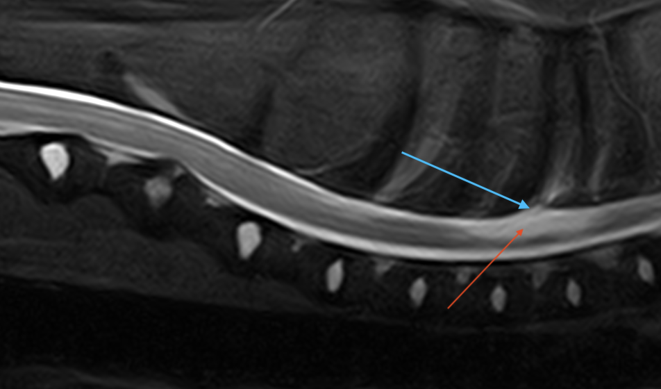
Fig 2 – A T2W (A) and T1W (B) transverse image of the vertebral column at the level of T3. The bifid nature of the spinous process can be noted on these images (blue arrows) with a linear soft tissue tract leading from the cutaneous structures (red arrow) ventrally between the osseous processes.
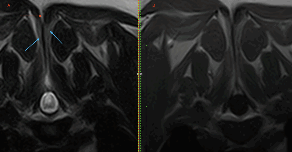
Fig 3 – A T2W (A) and T1W (B) transverse image of the vertebral column at the level of T3-T3 disc space. The intramedullary lesion noted in Fig. 1 can be seen to occupy approximately 75% of the cross-sectional area of the cord (blue arrow). This is compatible with a syrinx (syringomyelia). The dorsal subarachnoid space has lost its integrity and protrudes with the underlying neural tissue dorsally through the cleft in the dorsal lamina (blue oval).
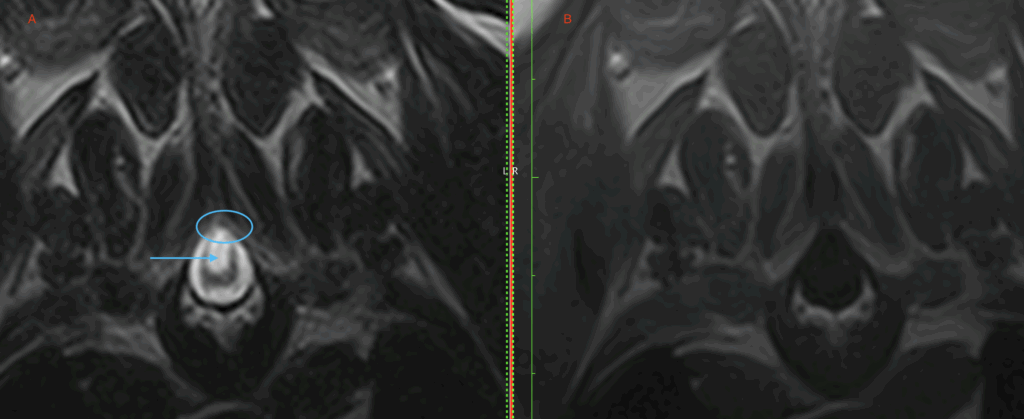
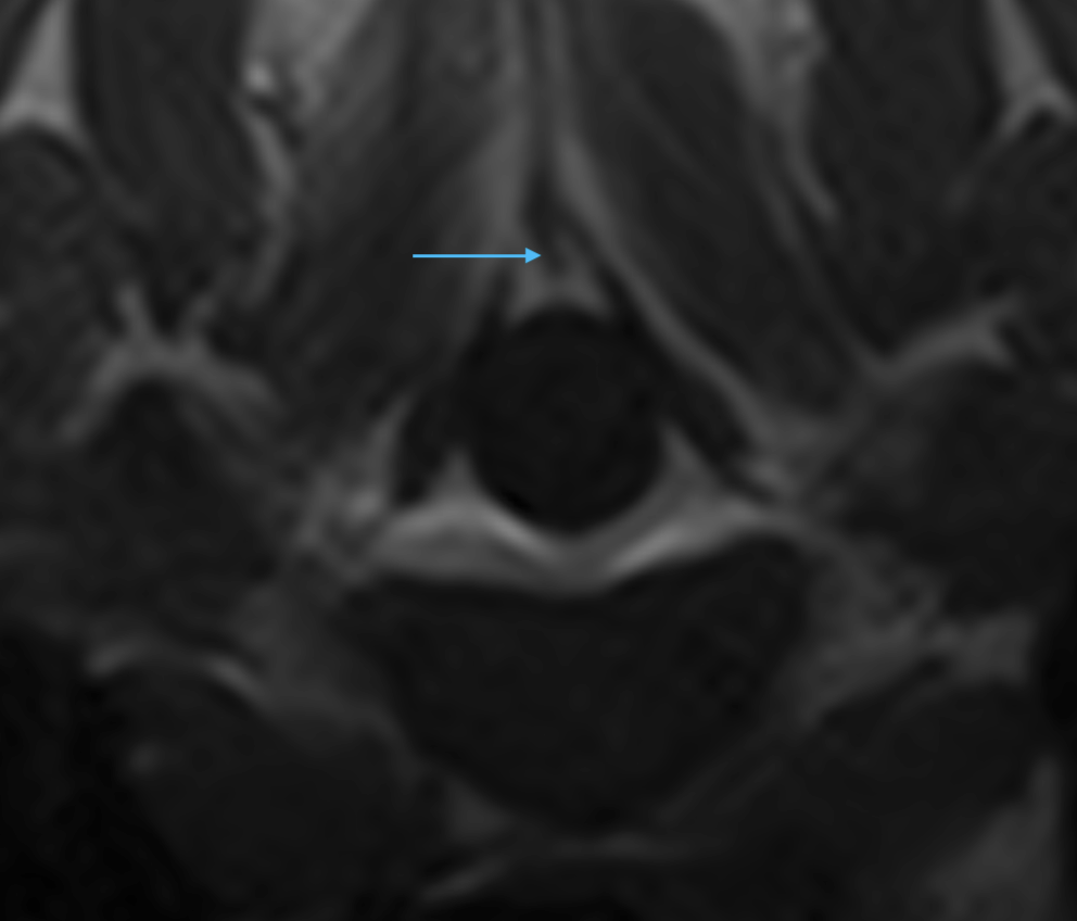
Fig 4 – A T1W transverse image of the vertebral column at the level of T3-T4. A linear soft tissue tract can be seen attached to the meninges and extending dorsally through the osseous cleft in the lamina (blue arrow).
Based on the images, surgical resection of the sinus tract was performed along with a dorsal laminectomy at T3-T4 to allow access to the dura mater. Descriptions of the surgical approach can be found in the references below. The dog recovered without any complications, and at her 2-week post-operative recheck, was assessed to be stable.
Disease overview
A dermoid sinus is defined as a congenital malformation resulting from a failed separation of the neural tube from the skin (ectoderm). The result is typically a tubular sac that is lined with hair follicles, sweat glands, and sebaceous glands, extending from the dorsal midline to underlying tissues. Six different types have been described, depending on the ventral extent of the tubular sac. According to this classification, if the open sinus tract reaches downward to the level of the dura mater it is classed as a type IV, as seen in this case.
Several reports have documented the presence of a dermoid sinus in association with various skeletal conditions, including hemivertebra and spina bifida in dogs and cats. Surgical treatment may not be required if the sinus is not responsible for clinical signs and is not connected to the dura mater. Cases with neurological deficits without treatment carry a guarded prognosis, as neurological function can continue to deteriorate because of further inflammation, infection, or cord compression due to the accumulation of hair debris. A dorsal laminectomy is usually necessary to completely excise the sinus and break down the adhesions with the dura mater.
Spina bifida is a congenital (dorsal) cleft of the spinal column with or without protrusion of the meninges and sometimes the spinal cord. A meningocele is a protrusion of meninges through a laminar defect in the spinal column, forming a dural sac filled with cerebrospinal fluid and a meningomyelocele is a protrusion of meninges and spinal cord or neural tissue through a defect in the spinal column. The clinical suspicion requires confirmation with CT or MRI. Although CT can assist with the 3-dimensional understanding of the bony defect, the neural and soft tissue defect requires MR imaging to be identified, especially if resection surgery is to be considered.
With many thanks to Dr. Carrie Jurney and the team at Remedy Veterinary Specialists, San Francisco, CA, for providing this study.
References
Kopke MA, Jack MW, Baltzer WI, Wightman PF, Gal A. Dermoid sinus type VI associated with spina bifida and tethered cord syndrome in a French Bulldog. J Vet Diagn Invest. 2019 Mar;31(2):294-297.
Song RB, Glass EN, Kent M. Spina Bifida, Meningomyelocele, and Meningocele. Vet Clin North Am Small Anim Pract. 2016 Mar;46(2):327-45
Ployart S, Doran I, Bomassi E, Bille C, Libermann S. Myelomeningocoele and a dermoid sinus-like lesion in a French bulldog. Can Vet J. 2013 Dec;54(12):1133-6


