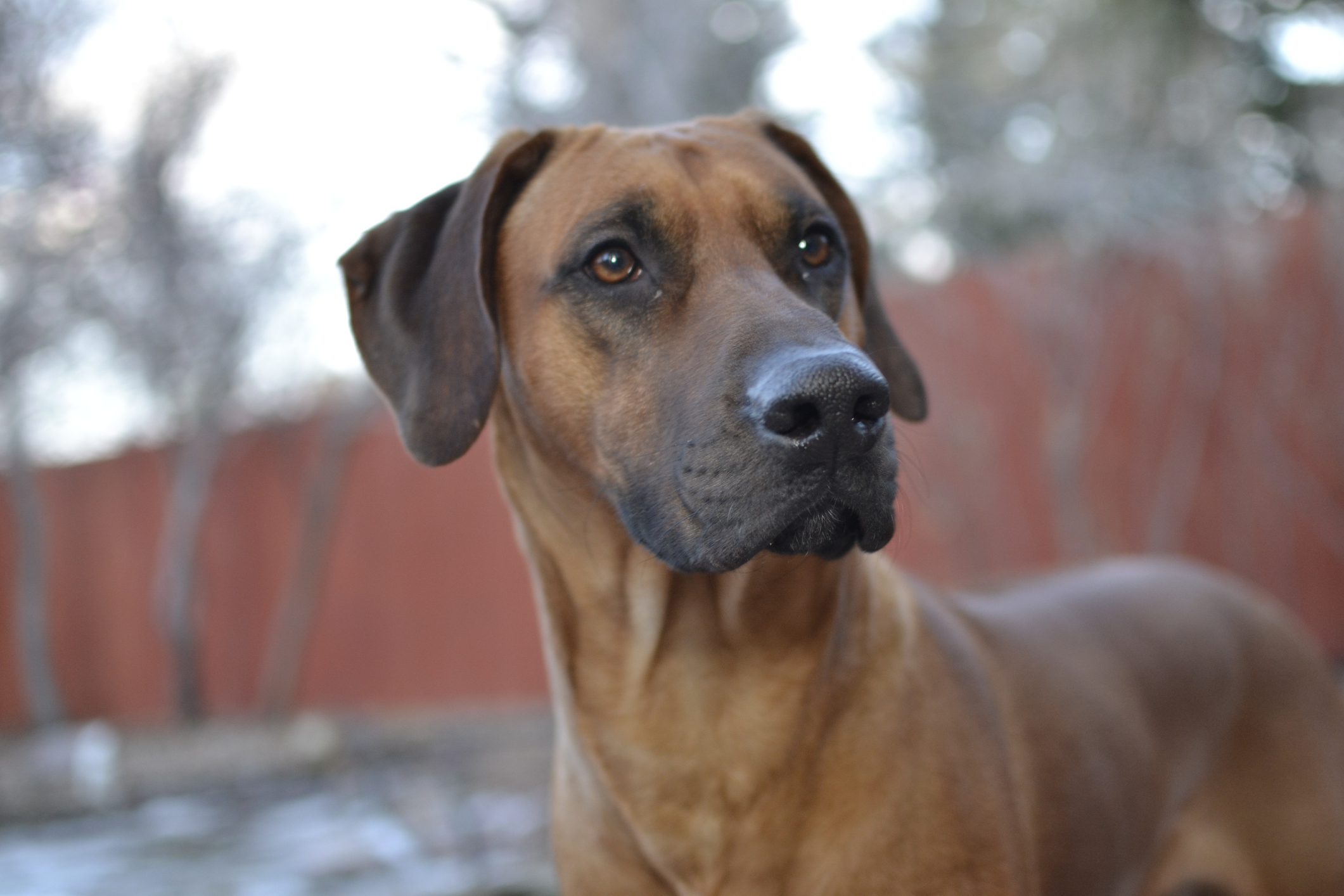



MRI is the imaging modality of choice for dermoid sinus, offering superior soft-tissue resolution for mapping sinus tracts and assessing intracranial or intraspinal extension. In this case study, we take a look at a dermoid sinus in a young Rhodesian Ridgeback.
The patient
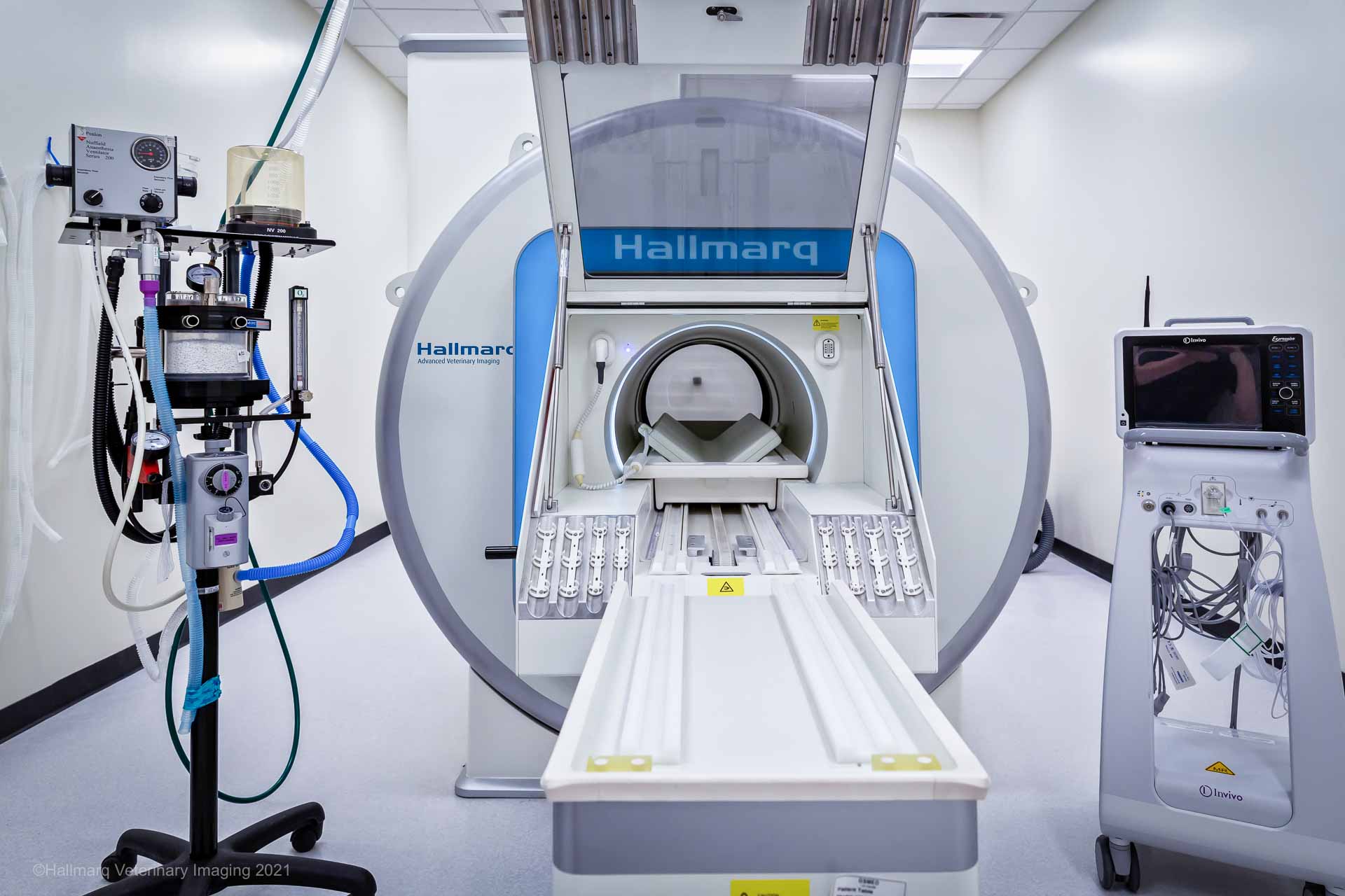
A 1-year-old female spayed Rhodesian Ridgeback presented with a several-month history of a recurrent draining tract on the dorsal cervical region. Otherwise considered healthy, the dog underwent a thoracolumbar vertebral column MRI using the Hallmarq third generation 1.5T machine (see Figs 1 and 2).
The imaging results
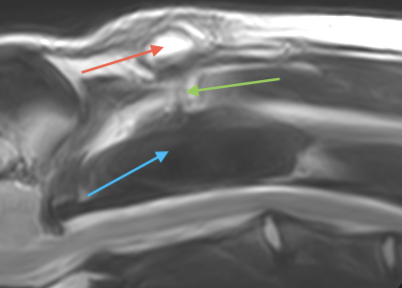
Fig 1 (A) below: shows a T2W sagittal image of the cranial cervical vertebral column. There is an ill-defined and irregular multilobulated subcutaneous structure (red arrow), with a more linear structure (green arrow) extending toward the spinous process of the C2 vertebra (blue arrow). The subcutaneous lesion is heterogeneously T2W hyperintense to muscle with a more isointense component of the linear structure.
In Fig 1 (B), the lesion can be noted to be more isointense in the top image (T1W pre-contrast) with strong heterogeneous contrast enhancement on the bottom image (T1W post-contrast).
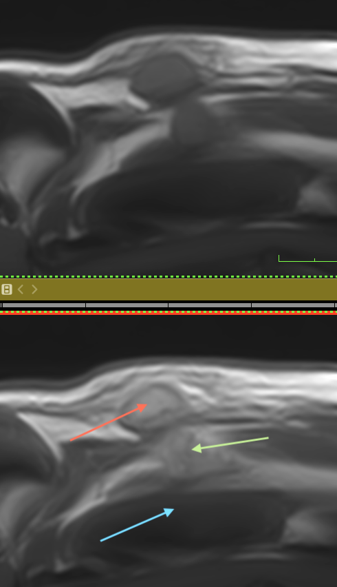
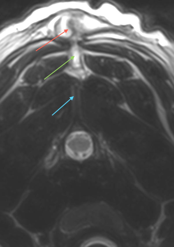
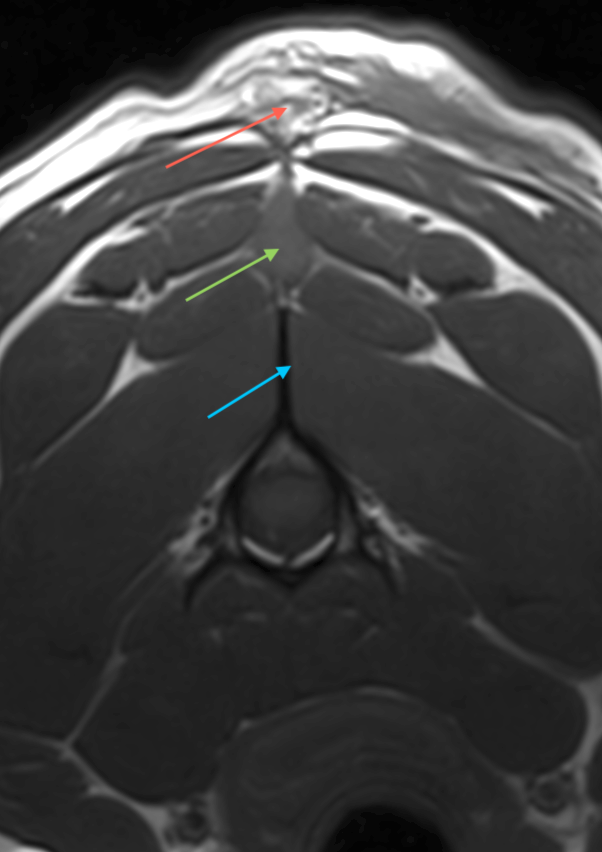
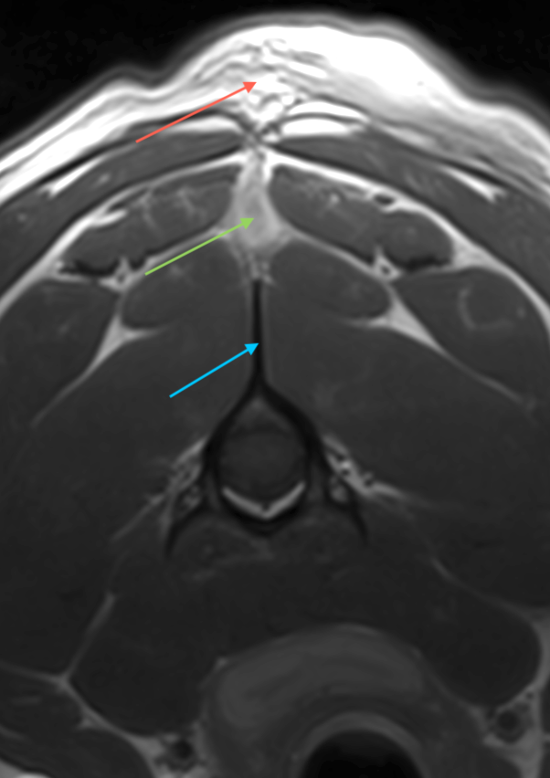
Fig 2 – A T2W (A), T1W (B) and T1W post-contrast (C) transverse image of the vertebral column at the level of C2. There is a relatively well-defined, ovoid subcutaneous mass (red arrow) with a linear ventral extension (green arrow) noted between this structure and the spinous process of C2 (blue arrow).
Dermoid sinus: the pathology
Based on the images, surgical resection of the sinus tract was performed. Descriptions of the surgical approach can be found in the references below. Happily, the dog recovered without any complications. Based on histopathology of the excised tissue, a focally extensive region of granulomatous inflammation centered around loose hair shafts and bordered by dense fibrous connective tissue was seen within the dermis and subcutis. In one of the tissue sections, a cyst was noted, lined by stratified squamous epithelium and containing lamellar keratin and fragments of hair shafts.
There were well-developed small hair follicles and apocrine glands in the collagen surrounding the cyst. The findings were compatible with a ruptured dermoid sinus with regional granulomatous dermatitis and panniculitis.
Dermoid sinus: a disease overview
Dermoid sinus (DS) is a congenital malformation arising from incomplete separation of the ectoderm from the neural tube during embryogenesis. As a result, this produces a tubular tract along the dorsal midline that can extend from the skin surface to the spinal cord. The condition is most prevalent in Rhodesian Ridgebacks, where breed-wide prevalence reaches approximately 2.53%.
The dorsal hair ridge characteristic of Rhodesian Ridgebacks is caused by a 133-kb duplication involving four fibroblast growth factor genes (FGF3, FGF4, FGF19, ORAOV1), and this same dominant mutation predisposes dogs to dermoid sinus. FGF dysregulation during embryonic development is the proposed mechanism linking the ridge phenotype to the sinus defect.
DS lesions are most commonly located in the cervical region, followed by thoracic and sacrococcygeal sites, and present as skin openings or subcutaneous swellings on the dorsal midline. Lesions are classified by depth of penetration:
- Type I: attachment to or ending at the supraspinous ligament
- Type II: connected to the supraspinous ligament via a fibrous strand
- Type III: extends deeper without dural communication; may require laminectomy
- Type IV: penetrates the spinal cord or dura mater
Neurological signs are absent in most cases but can arise from infection disseminating to the spinal cord or from direct cord tethering, particularly in Type IV lesions.
Why MRI for dermoid sinus?
MRI is the imaging modality of choice for dermoid sinus, offering superior soft-tissue resolution for mapping sinus tracts and assessing intracranial or intraspinal extension. Typical findings include fluid-filled structures, fibrous capsules, and sinus tracts, though signal characteristics vary with cyst content. MRI reliably identifies fluid-filled structures, fibrous capsules, and sinus tracts but often cannot identify the termination of the tract. However, conventional MRI may not adequately show sinus tracts if they are small or fall outside the imaging plane.
- 3D MR myelography can demonstrate communication between a dorsal dermal sinus and the intraspinal space when conventional MRI cannot
- Fat-suppressed T1 with gadolinium highlights infectious complications
In previously described Rhodesian Ridgeback dogs, MRI of type I dermoid sinuses identified fluid-filled structures and fibrous capsules but could not determine tract termination, similar to the case described here.
Surgical excision is the treatment of choice and is curative in most cases. Complete resection up to the spinous process or supraspinous ligament is the standard approach. In addition, with partial dorsal laminectomy is required when the sinus communicates with the vertebral canal or spinal cord. Postoperative wound healing is typically uncomplicated, with no recurrence reported at 6-month follow-up in adequately resected cases.
With thanks to Dr. Adam Moeser and team at Wisconsin Veterinary Neurology & Surgical Center for sharing this study with us.
References
- Distl, O. (2022). Prevalence and segregation analysis of dermoid sinus in Rhodesian Ridgebacks.. Veterinary journal, 105803 . https://doi.org/10.1016/j.tvjl.2022.105803
- Hillbertz, N. S., & Andersson, G. (2004). P‐37 Dermoid sinus in Rhodesian ridgeback dogs. Veterinary Dermatology, 15, 52-52. https://doi.org/10.1111/j.1365-3164.2004.00414_37.x
- Hillbertz, N. S., Isaksson, M., Karlsson, E. K., Hellmén, E., Pielberg, G., Savolainen, P., Wade, C., Euler, H., Gustafson, U., Hedhammar, Å., Nilsson, M., Lindblad-Toh, K., Andersson, L., & Andersson, G. (2007). Duplication of FGF3, FGF4, FGF19 and ORAOV1 causes hair ridge and predisposition to dermoid sinus in Ridgeback dogs. Nature Genetics, 39, 1318-1320. https://doi.org/10.1038/ng.2007.4
- Rahal, S., Mortari, A., Yamashita, S., Filho, M. M., Hatschbac, E., & Sequeira, J. (2008). Magnetic resonance imaging in the diagnosis of type 1 dermoid sinus in two Rhodesian ridgeback dogs.. The Canadian veterinary journal, 49 9, 871-6 .