
In this case study, a thoroughbred racehorse was presented with acute left forelimb lameness following exercise. A fetlock fracture was suspected and radiographic images highlighted several areas of concern.
Radiographic findings
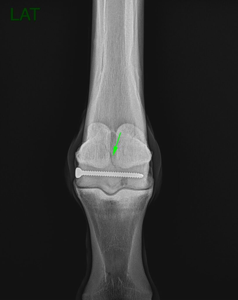
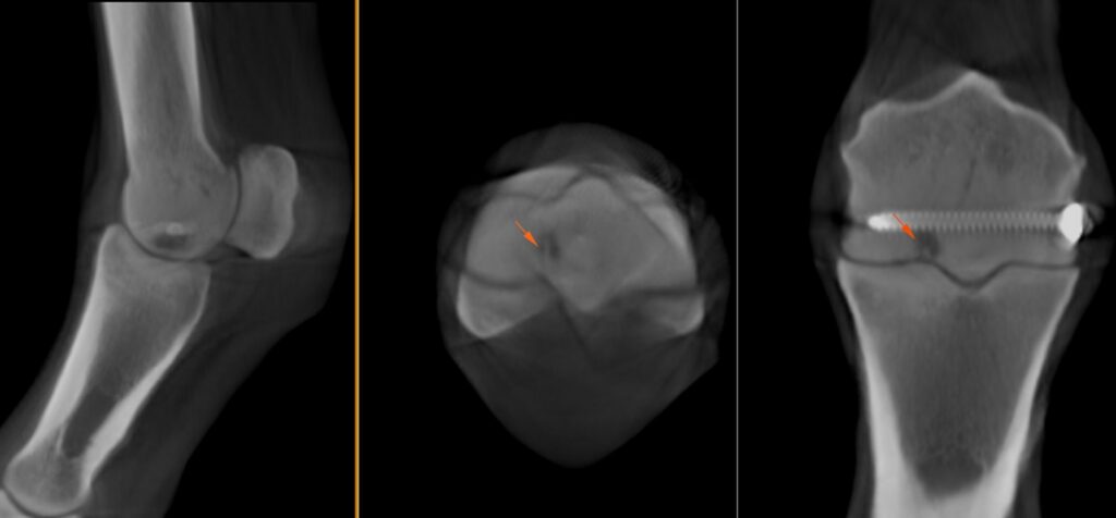
The primary concern identified in the radiographs was a faint line at the proximal aspect of the sagittal ridge, potentially indicating a fracture line (Figure 1). Additionally, an area of osteolysis was observed at the medial parasagittal groove of the medial condyle of the third metacarpus. This osteolytic area was most likely in communication with the joint. There was also a surgical implant (screw).
CT findings
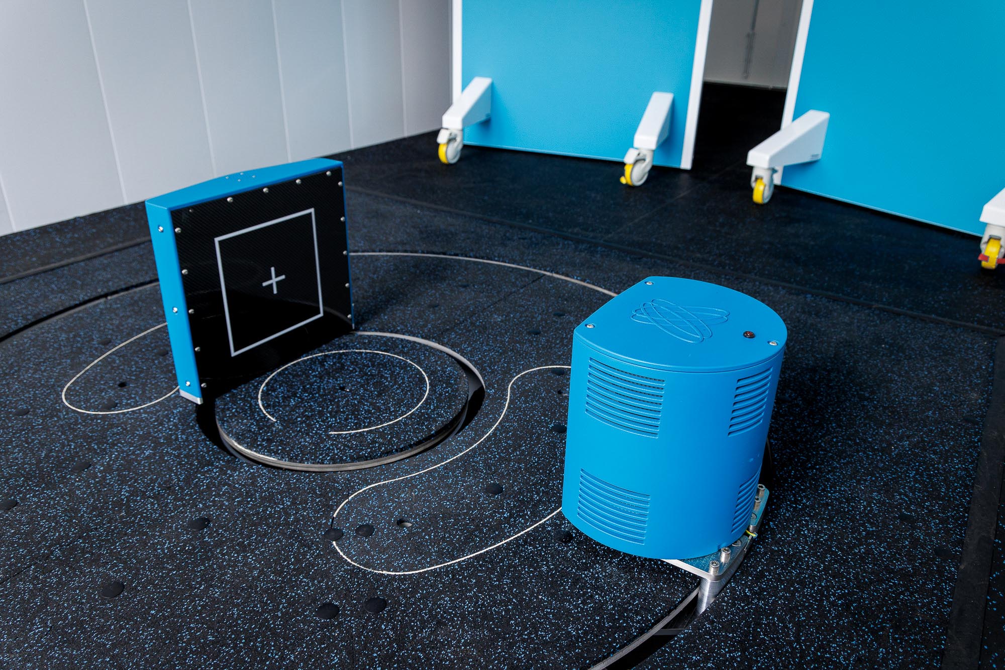
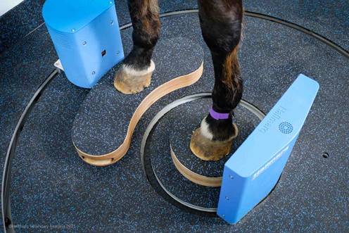
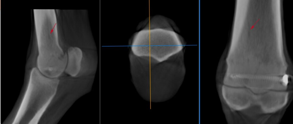
With a fetlock fracture suspected, the horse underwent a scan using Hallmarq’s Vision CT machine. This revealed several hypoattenuating lines entering the palmar metacarpus at the level of the physeal scar abaxial to the proximal aspect of the sagittal ridge of the third metacarpus that likely represented a course of blood vessels (Figure 2).
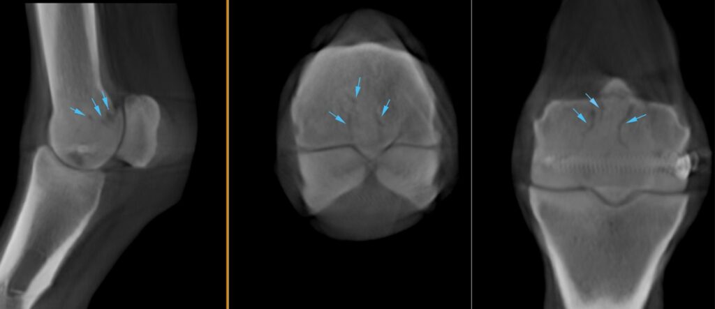
The area of osteolysis at the parasagittal groove of the medial condyle was well visualised, showing direct communication with the fetlock joint though two tracts (Figure 3). Additionally, the CT revealed a hypoattenuating line visible on all planes within the distal diaphysis of the third metacarpus extending from the region of the medial parasagittal groove spiralling medially, likely indicating a hair line fracture (Figure 4).
Conclusion
Despite the artefacts caused by the metal screw, assessing for fetlock fracture with a CT scan allowed for an efficient and detailed evaluation, something that would have been impossible with MRI due to metal artefact. The initial suspicion of a fetlock fracture was dispelled however, revealing that the observed structures were blood vessels. However, the CT scan identified a second area of potential fracture. As a result, ongoing monitoring and a follow-up CT scan in a few weeks were recommended.
This case was kindly provided by Henry O’Neil MVB, DVM, MS, Dipl ACVS, MRCVS, Donnington Grove Equine Vets, UK.