

Canine hydranencephaly
The patient
We look at the clinical study of a 1-year-old male mix-breed dog who presented with intermittent circling, a reduced level of consciousness, and intermittent confusion, in addition to weight loss. A neurological examination confirmed an asymmetrical forebrain lesion. The dog underwent an MRI scan using the Hallmarq small animal 1.5T machine.
The imaging results
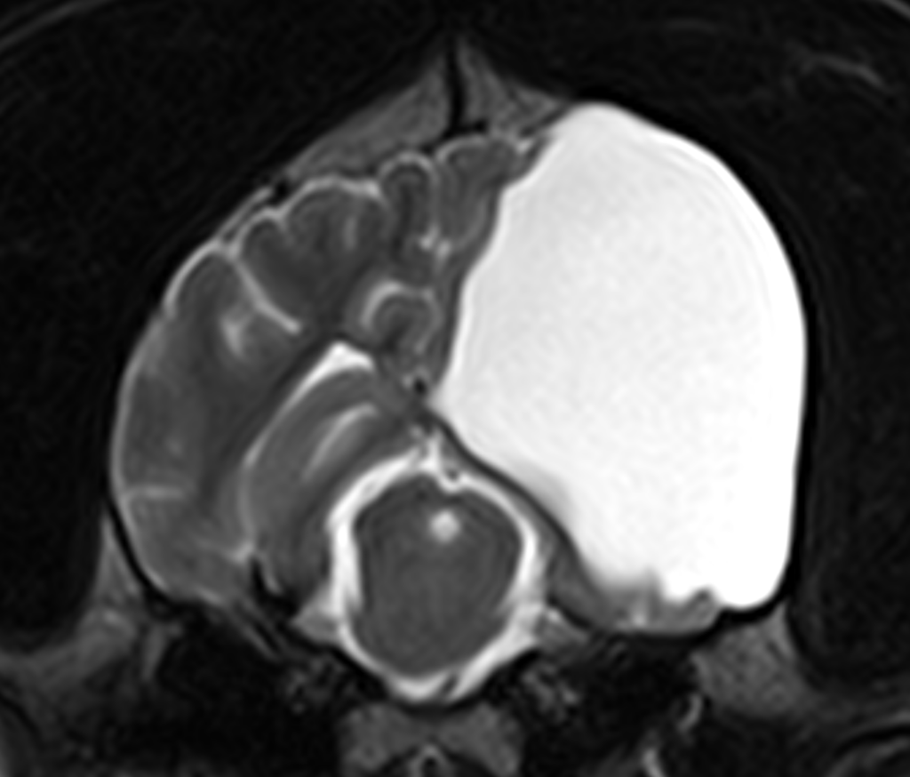
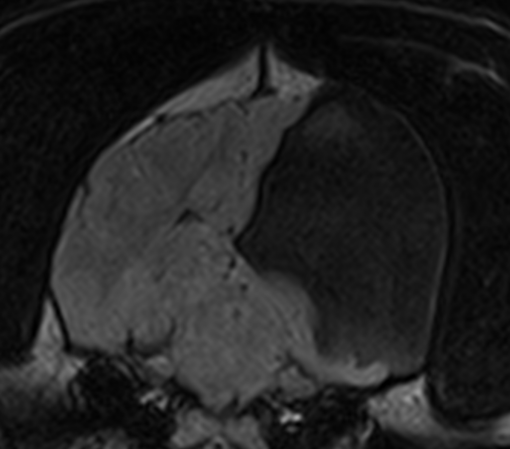
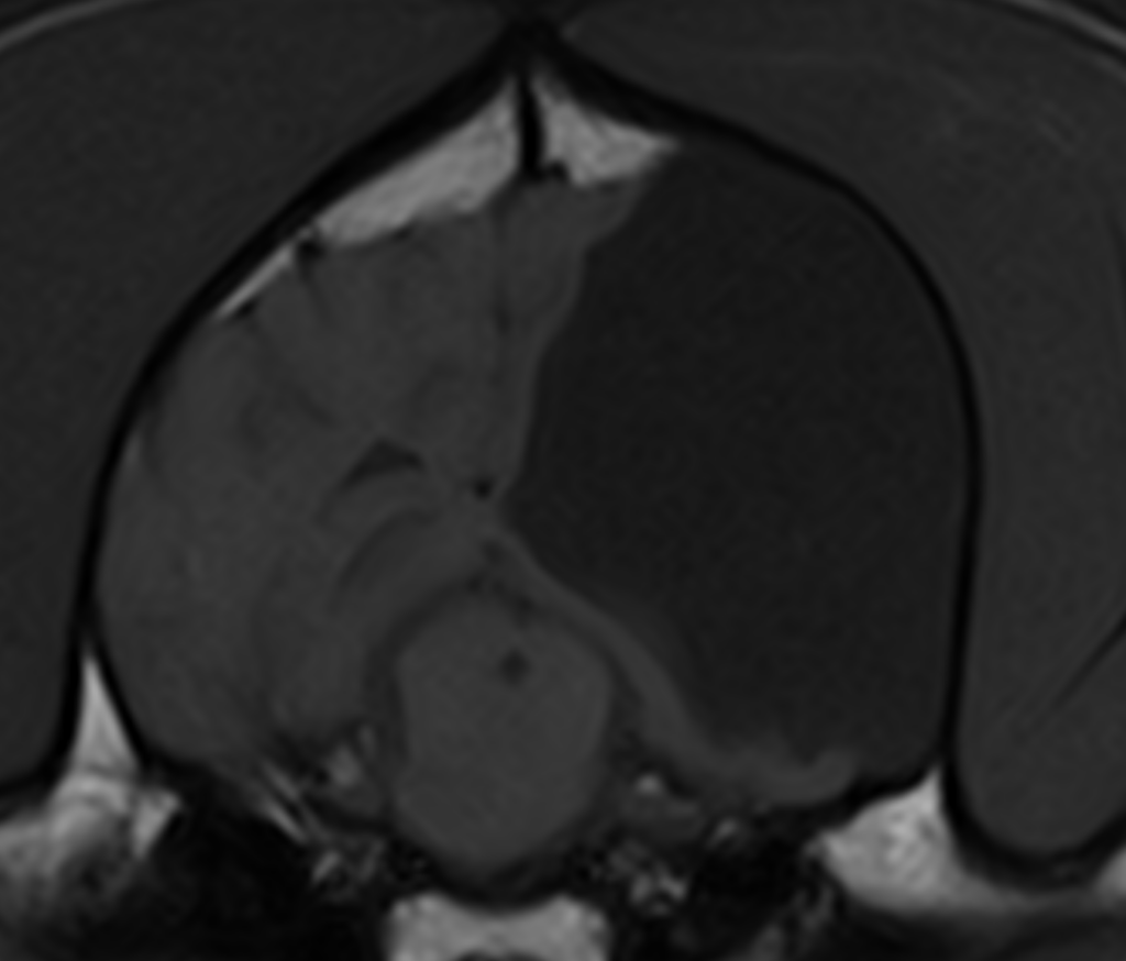
A large fluid-filled lesion is associated with an almost complete absence of the frontal, temporoparietal, and occipital lobes on the left side (A). The contents of this lesion are isointense to CSF on all sequences and suppress on T2-FLAIR (B); the lesion is hypointense on T1W and does not enhance post-contrast. This lesion is associated with mild calvarial malformation on that side resulting in an asymmetrical and larger cranial vault on the affected side; this is presumed to be an associated pressure expansion of the left calvaria with a dome-shaped expansion of the diploe (C). On the left side, there is diminished thalamic mass (D). Very mild mass effect is associated with this lesion with a subtle midline shift to the right. The imaging study is compatible with a diagnosis of hydranencephaly.
The most valuable MRI sequence for this case
The combination of the image sequences in this case allows for confirmation of the lesion contents to be compatible with CSF while allowing for an assessment of the overlying bone. If the contents had not been CSF, they would likely have a different intensity to that seen within the normal contralateral ventricle. Get in touch with our team to find out more about Hallmarq’s veterinary-specific sequences:
Disease overview
Hydranencephaly is an abnormality in which the cerebrum is destroyed in utero and replaced by a CSF-filled cavity without residual neural parenchyma. A similar malformation is porencephaly, a CSF-filled cavity within the brain that may communicate with the ventricular system or subarachnoid space and is a more focal and less extensive defect compared with hydranencephaly.
While hydranencephaly may present in puppies with compulsive and aggressive behaviour which may or may not be progressive, porencephaly should be considered as a differential in adult dogs presenting with seizures. Vestibular signs, visual dysfunction, and altered mentation and consciousness have also been described. Hydranencephaly and porencephaly are caused by fetal or perinatal brain destruction, possibly secondary to trauma.
Why advanced imaging for hydranencephaly?
Advanced imaging is necessary to define the structural abnormalities associated with porencephaly and hydrancephaly. MRI shows a variable loss of cerebral hemisphere parenchyma, resulting in cavitary lesions that communicate with the ipsilateral lateral ventricle. The content of the cavity is isointense to the CSF on all image sequences and is suppressed on FLAIR images. Most dogs have unilateral lesions and the extent of parenchymal loss varies between dogs. Dogs can also have asymmetry of the crus cerebri, transverse and longitudinal fibres of the pons and the pyramids with the smaller side being ipsilateral to the side of the cavitary lesion. CSF analysis should be within normal limits.
Ventriculoperitoneal shunt placement may ameliorate the clinical signs. Similar medical treatment to that used for hydrocephalus can be considered with the institution of anti-inflammatory corticosteroids, omeprazole, and even topiramate. Prognosis will be determined by the extent of the cavitation and the response of the clinical signs to symptomatic therapy or surgery if deemed appropriate.
Our thanks to Dr. Amanda Full DVM, DACVIM at Premier Veterinary Group, Chicago for sharing this study.
References
- Davies ES, Volk HA, Behr S, Summers B, de Lahunta A, Syme H, et al. . Porencephaly and hydranencephaly in six dogs. Vet Rec. (2012) 170:179
- Davini T, Mattei C, La Rosa C, Remelli C, Specchi S, Lionello E, Dell’Era E, Bernardini M. Are postnatal traumatic events an underestimated cause of porencephalic lesions in dogs and cats? Front Vet Sci. 2023 Dec 6;10:1302399
- Schmidt MJ, Klumpp S, Amort K, Jawinski S, Kramer M. Porencephaly in dogs and cats: magnetic resonance imaging findings and clinical signs. Vet Radiol Ultrasound. 2012 Mar-Apr;53(2):142-9.


