

In this case study, we take a look at chronic solar penetration and how the use of both Standing Equine MRI and Vision CT – combined – helped with a definitive diagnosis that was truly life-saving for the horse.
Case history
The horse had a 4-week history of a puncture wound to the right forefoot, caused by a nail penetration through the frog. The nail was removed on the farm, and the horse was managed conservatively. Despite this, the horse clinically worsened. On admission to the clinic the horse was grade 5/5 lame in the right forelimb and draining tracts were present at the frog and heel regions, with pronounced inflammation of the surrounding soft tissues.
MRI findings
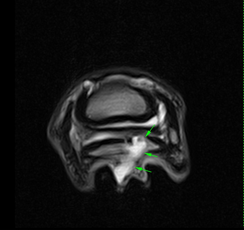
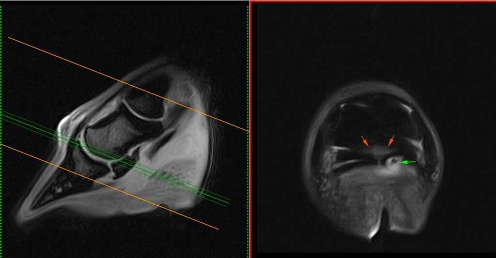
Susceptibility artefact was seen at the lateral sulcus of the frog. A wide area of hyperintense STIR and T2 signal, and hypointense T1 signal, extended from the lateral sulcus axially through the medial lobe of the DDFT (Fig. 1), terminating at the proximal aspect of the flexor surface of the navicular bone. Hyperintense STIR signal was present at the medial and axial aspect of the navicular bone, consistent with fluid signal (Fig. 2). The navicular bursa and distal interphalangeal joint were not effused.
A full-thickness tear of the DDFT was visualised; however, the flexor surface of the navicular bone could not be easily assessed, apart from the marked fluid signal present, which could correspond to septic osteitis.
CT findings
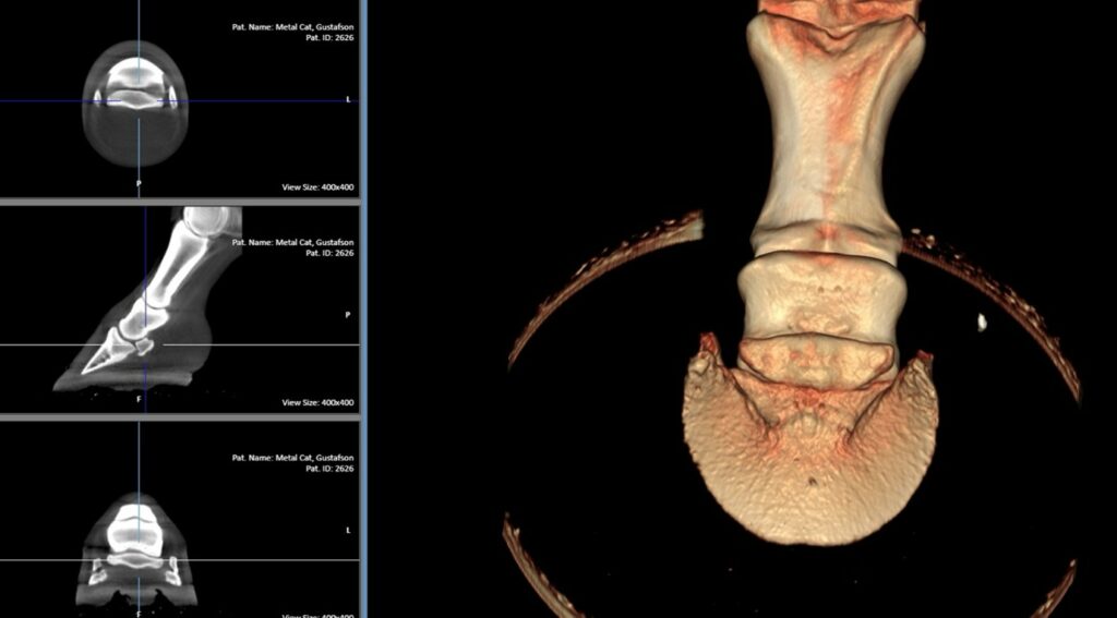
A CT scan using Hallmarq’s Vision CT provided clear visualisation of the flexor surface of the navicular bone (Fig. 3). There were no signs of septic osteitis or osteomyelitis of the bone on CT, despite the concerning MRI findings. CT 3D reconstruction was visually helpful to the surgical team in planning the surgical approach and in avoiding damage to the osseous structures.
The outcome
The horse underwent what is known as the street nail procedure. The frog was incised along the puncture tract, which was then followed to its deepest point, potentially involving the navicular bursa, distal interphalangeal joint, or deep digital flexor tendon sheath. Any foreign material, necrotic tissue, or debris was removed, and any infected synovial structures were thoroughly lavaged. At discharge the horse was walking sound and showing progressive improvements with a fair to good prognosis given.
At the most recent follow-up, the horse continues to improve and is doing well with minimal lameness, and I anticipate he will return to soundness.
Conclusion
For many years, Standing Equine MRI has been regarded as having superior diagnostic capability for solar penetrating injuries, as it significantly aids diagnosis and treatment planning without the risks associated with general anaesthesia (1-6). The advantages of combining CT and MRI for imaging of the distal limb have been recently documented (7), and this case serves as a strong example.
Without access to combined CT and MRI, cases such as this may previously have been given a grave prognosis at admission and not considered surgical candidates, due to the long-standing infection and presumptive involvement of both bone and soft tissue. In this instance, these imaging modalities were truly life-saving for the horse, in combination with the expertise of our surgical team led by Dr Cailey Nichols.
With thanks to Dr. Chris Bell, Elders Equine Clinic, Canada, for sharing this case with us.
References
1.URRACA DEL JUNCO, C.I., Mair, T.S., Powell, S.E., Milner, P.I., Font, A.F., Schwarz, T. and Weaver, M.P., 2012. Magnetic resonance imaging findings of equine solar penetration wounds. Veterinary Radiology & Ultrasound, 53(1), pp.71-75.
2.De Heer, N., Compagnie, E. and Ter Braake, F., 2015. Penetrating solar wounds to the foot: benefit of MRI in treatment decisions. Vlaams Diergeneeskundig Tijdschrift, 84(1).
3.Dyson, S. and Murray, R., 2010. The foot and pastern. Equine MRI, pp.269-314.
4.Sherlock, C.E., Kinns, J. and Mair, T.S., 2007. Evaluation of foot pain in the standing horse by magnetic resonance imaging. Veterinary Record, 161(22), pp.739-744.
5.Findley, J.A., Pinchbeck, G.L., Milner, P.I., Bladon, B.M., Boswell, J., Mair, T.S., Suthers, J.M. and Singer, E.R., 2014. Outcome of horses with synovial structure involvement following solar foot penetrations in four UK veterinary hospitals: 95 cases. Equine veterinary journal, 46(3), pp.352-357.
6.Scharf, A. and Peroni, J., 2025. Diagnostic imaging and management of penetrating wounds of the foot. Equine Veterinary Education
7.Taylor, S., Kelly, P., Daniel, C., James, O., McMaster, M. and Schwarz, T., 2025. Comparison of cone beam CT and low-field MRI of the distal limb of 85 standing sedated horses. Authorea. May 19, 2025


