

Metastatic intracranial choroid plexus tumours
The patient
A 7-and-a-half-year-old male neutered Pitbull presented for an onset of recurrent seizure and status epilepticus. The dog had been treated in hospital with a constant rate infusion of midazolam, intravenous phenobarbital, and intravenous levetiracetam. The seizures were effectively controlled. Otherwise considered healthy, the dog underwent a brain MRI using the Hallmarq 1.5T unit (see Fig 1).
The imaging results


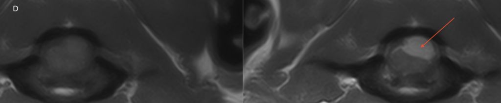
Red arrow – enhanced mass lesion within the 4th ventricle at the level of the choroid plexus
Red arrow – enhanced intradural mass lesion dorsal to the cord
There is a relatively well-defined ovoid extra-parenchymal mass lesion centered on the right-sided choroid-plexus of the 4th ventricle. The lesion is T2W and T1W iso to mildly hyperintense to grey matter with strong and homogenous contrast enhancement with extension along the regional meninges. There are no T2* signal voids within the lesion.
There are 3 other intradural extra-parenchymal masses with similar signal characteristics: these are seen on the left dorsolateral aspect of the spinal cord at C1, on the right ventrolateral aspect of the spinal cord at the C1-2 junction, and on the right ventrolateral aspect of the spinal cord over the cranial C2 vertebral body.
There is moderate distension of the lateral ventricles and the olfactory recesses, with periventricular T2W hyperintensity. There is mild regional parenchymal compression associated with the masses.
The main differentials for these lesions include choroid plexus tumour with ‘drop’ metastases, multiple meningioma, histiocytic sarcoma, and lymphoma.
The most valuable MRI sequence for this case
The T1W post-contrast sequence demonstrates both meningeal involvement and the homogenous enhancement often seen associated with tumours such as choroid plexus tumors and meningiomas originating outside of the blood-brain barrier. Typically, the tissue that enhances defines the extent of the disease.
Disease overview
Choroid plexus tumors (CPT) account for approximately 7–10% of the central nervous system (CNS) neoplasia in dogs and affect the fourth ventricle in 46% of CPTs. The current classification recognizes three grades of CPTs: choroid plexus papilloma (CPP, grade I), atypical CPP (aCPP, grade II), and choroid plexus carcinoma (CPC, grade III).
In a clinical setting, differentiation between CPP and CPC involves CSF analysis and magnetic resonance imaging (MRI) features. A CSF total protein concentration above 80 mg/L and MRI presence of CSF drop metastases are strongly suggestive of CPC, with 33% exhibiting subarachnoid metastases while their presence is considered rare with CPP.
CSF drop metastases refer to the metastatic spreading of a primary CNS neoplasia to the subarachnoid space and ventricular system of the CNS, following the CSF flow.
With thanks to Dr. Baye Williamson DVM, DACVIM (Neurology) at Veterinary Emergency + Referral Center (VERC), Hawaii, for providing this case study.
References
- Albertini GM, Malbon A, Staudacher A, Stabile F. Clinical, magnetic resonance imaging, and histological description of a choroid plexus papilloma with disseminated intraventricular and spinal cerebrospinal fluid drop metastases in a young adult dog: a case report. Front Vet Sci. 2023 Aug 3;10:1223729
- Westworth DR, Dickinson PJ, Vernau W, Johnson EG, Bollen AW, Kass PH, et al. Choroid plexus tumors in 56 dogs (1985-2007). J Vet Intern Med. (2008) 22:1157–65
- Yeamans CL, Gutierrez-Quintana R, Haley A, Lamm CG. Magnetic resonance imaging and clinical findings associated with choroid plexus spinal cord “drop” metastases. J Am Anim Hosp Assoc. (2017) 53:265–9


