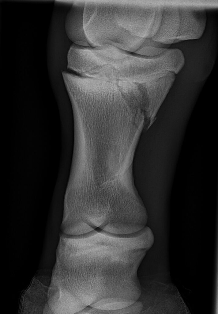
In this case study, a foal sustained a Salter-Harris type II fracture of the left hind proximal phalanx following a collision with a paddock mate, and subsequent fall. As a result, the foal was acutely lame on the affected limb. A radiographic examination confirmed the presence of the fracture, and the foal was referred to the hospital for further imaging and appropriate treatment.
CT findings
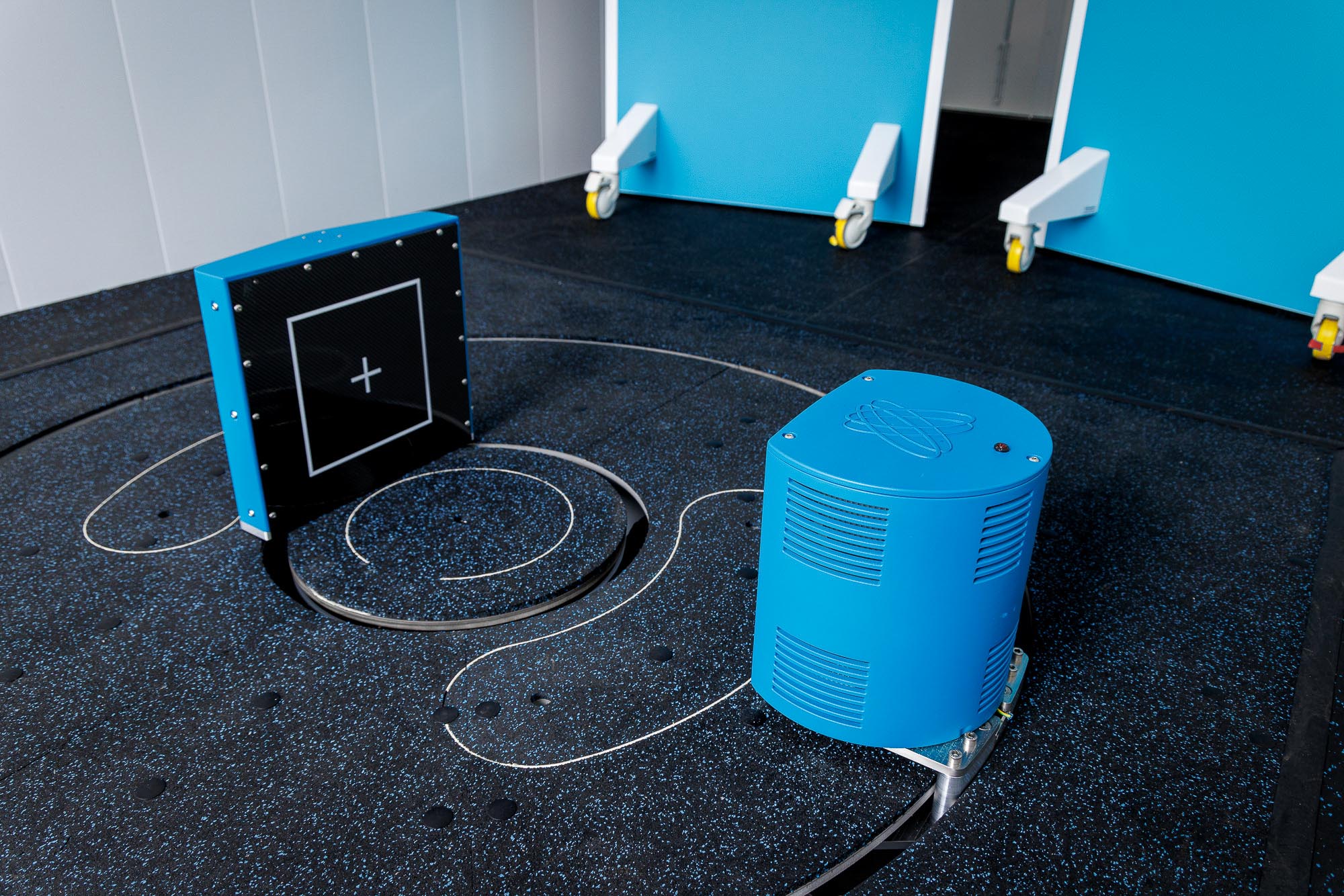
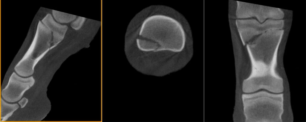
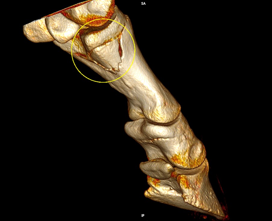
Upon arrival at hospital, the foal underwent computed tomography (CT) using Hallmarq’s Vision CT system. This confirmed a closed, displaced Salter–Harris type II fracture propagating through the physis from the lateral aspect, extending into the metaphysis on the plantaromedial aspect. In addition, multiple small osseous fragments were identified at the distal margin of the fracture fragment.
Treatment
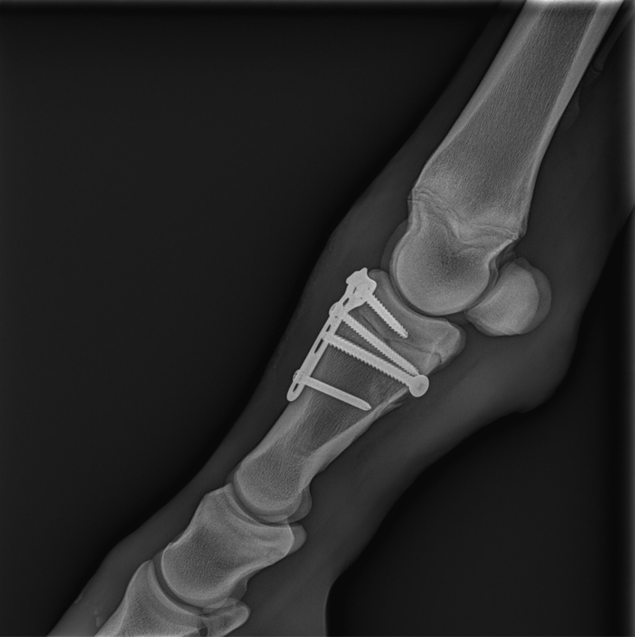
The following day, the fracture was surgically repaired under general anaesthesia using a combination of cortical screws and a plate. With fracture reduction achieved under intraoperative radiographic guidance, stabilisation was completed without complications. The foal was comfortable in the immediate postoperative period.
Conclusion
In the case of a Salter-Harris type II fracture, computed tomography (CT) allowed thorough evaluation and proved to be a suitable imaging modality. It enabled detailed assessment under mild sedation with the foal remaining standing, even at a young age. This facilitated accurate characterisation of the fracture configuration and supported informed surgical planning. In time, the case will be reviewed and follow-up information will be provided.
With thanks to surgeons Dr Henry O’ Neil and Dr Giorgio Ricardi, Donnington Grove Equine Vets, UK, for sharing this case with us.