Equine Veterinarians
Frequently asked questions
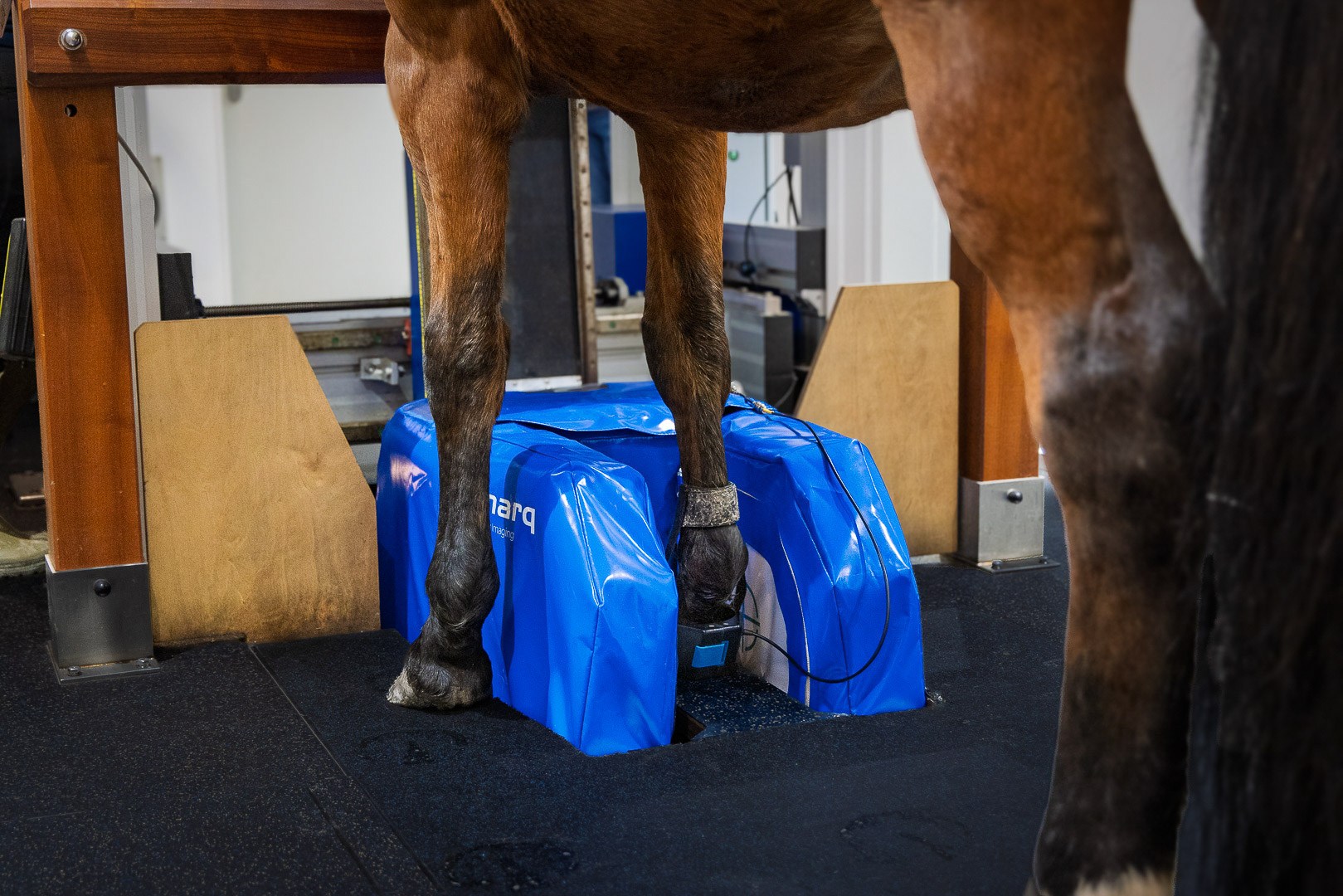
- Why refer for standing MRI?
- When would I refer for standing MRI?
- How do I refer for standing MRI?
- Why is MRI of the fetlock used for racehorses?
Much has been learned about the causes of equine lameness since the advent of MRI. From the previously under-diagnosed, such as collateral desmitis of the distal interphalangeal joint, through the previously misunderstood, such as navicular syndrome, to the previously unknown, such as bone marrow oedema, MRI has revolutionised our ability to provide a diagnosis and improve prognosis in equine lameness.
MRI is unparalleled in providing images of both soft and bony tissues. Distinguishing water from fat, it highlights areas of pathology such as inflammation and bruising, in a way that radiography, CT, ultrasound or nuclear scintigraphy just can’t do. By imaging the region of interest in slices orientated in any 3D plane, a lesion can be visualised without superimposition of adjacent structures. Multiple views allow you to appreciate the full extent of the injury.


