Distal limb injuries in horses are too often the result of stresses to both bony and soft tissue structures but Magnetic Resonance Imaging (MRI) can help aid diagnoses. Many breeds – in particular Western performance horses – experience extreme forces on the structures of the lower limb whilst executing transitions such as a tight turn or rapid acceleration.
Lameness subsequent to distal limb injury is common to both the performance and pleasure horse, resulting in time out of ridden work at best or career ending at worst. To learn more about the most common distal limb injuries in horses, continue reading.
Flexor Tendon Injury (Desmitis)
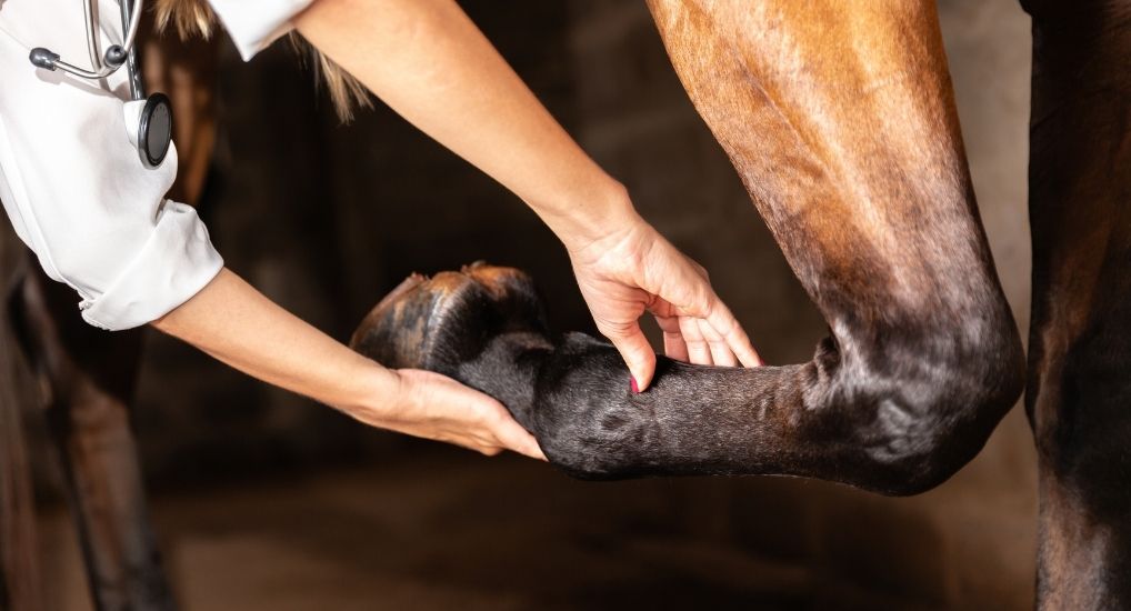
The classic bowed tendon, swelling, heat and associated lameness is usually easily recognized when a superficial or deep digital flexor tendon injury occurs above the foot. But what about when the injury is lower down the limb, within the hoof capsule? Assessment of more proximal tendon injuries can be performed with ultrasound but MRI is normally required to evaluate any damage occurring below the heel bulbs.
Fractures
Severity of distal limb fractures can range from mildly painful to catastrophic depending on location and type. Acute, severe lameness due to traumatic fracture can occur at any time, but repetitive loading during high intensity work and adaptive re-modelling can also result in catastrophic fracture. Early recognition of pre-cursor clinical indicators, appropriate diagnostics and adaptation of training can be preventative.
Ligament Injury
Suspensory ligament injury affecting the proximal third, body or branches is well-documented in competition and race horses. There may also be compounding factors such as fetlock joint involvement or foot imbalance. Suspensory ligament injury, injury to collateral ligaments stabilizing joints or ligaments within the hoof capsule can require detailed investigation to accurately diagnose the problem.
Laceration and Wounds
Traumatic injuries resulting in a laceration or wound can be the most dramatic in appearance, especially to an owner. Location, depth and involvement of any joint is important in determining likely severity, treatment requirements and prognosis
Penetrating Foreign Body Injuries
Seemingly innocuous, a small scab over dorsal carpus or a black tract on the sole of a foot may be the only indicator of serious injury. Determining depth and direction of a penetrating foreign body, for example a nail or clench in the sole of the foot, would help distinguish the difference between a sub solar abscess or a septic navicular bursa and damage to the deep digital flexor tendon.
Bony Concussive Trauma
Subchondral bone damage, often as a result of repetitive, concussive loading of the joint, can result in an insidious onset of persistent lameness. Diagnosis and monitoring via assessment of both structural and functional changes of the bone assists successful management of these cases.
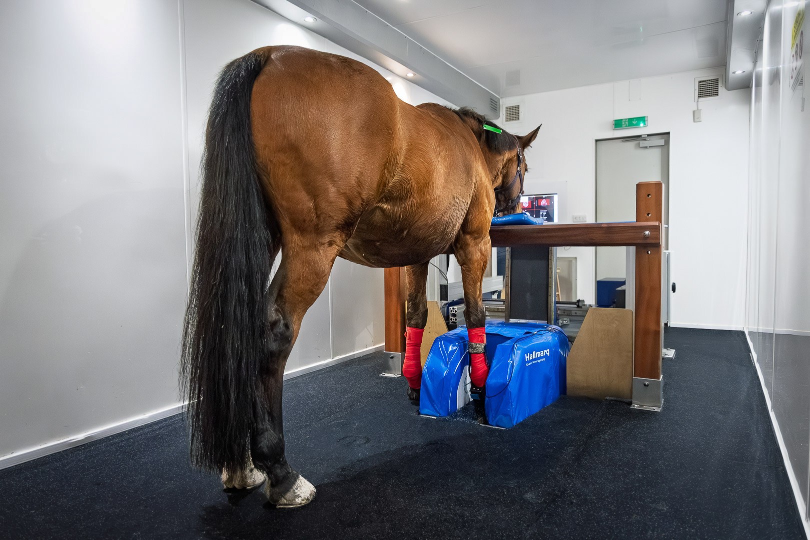
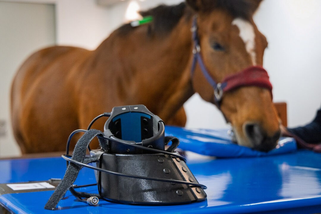
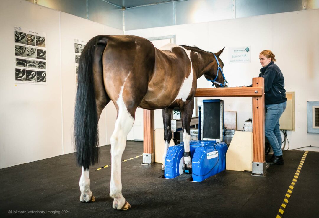
Getting a proper diagnosis of any of the most common distal limb injuries in horses will help equine health professionals work together to bring a horse back to competition health. Hallmarq Veterinary Imaging offers the latest technology, including standing equine MRI, to assist with accurate diagnosis for improved treatment planning and prognosis.
