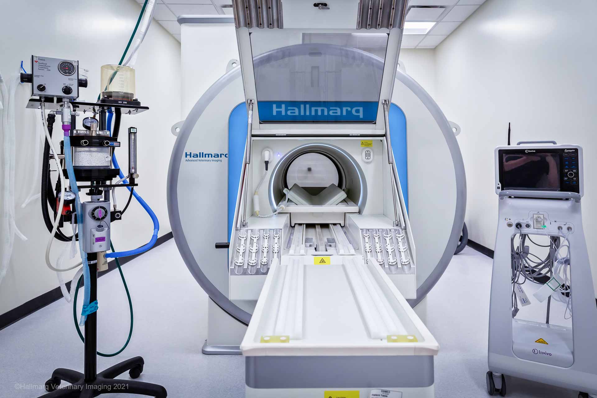
An MRI scan is an imaging technique that uses a strong magnetic field to generate highly detailed cross-sectional images of your pet’s internal structures. Unlike X-rays or CT scans, which are primarily useful for visualizing bones, MRI provides exceptional clarity when imaging soft tissues. This technology enables veterinary specialists to observe small details of the brain, spinal cord, ligaments, tendons and other soft tissue organs with unmatched precision.
MRI can confirm suspected diagnoses, exclude other potential causes of symptoms, precisely locate issues such as herniated discs or tiny brain tumors and identify disease locations if surgery is needed. Overall, an MRI scan is a valuable diagnostic tool that transforms veterinary medicine from educated guesses into well-informed decisions. The are invaluable when creating the most effective, targeted treatment plans for your pet.
Although MRI offers clear benefits, some misconceptions still surround this technology. This overview addresses five common concerns, distinguishing myths from facts, to help you make the best health decisions for your pet.
Myth 1 – MRI is Dangerous for Small Animals
The Concern: Many worry that the process is intimidating. Scenarios like anesthesia and a tube in a large magnet fostering fears that the procedure could be risky for small animals.
The Reality: Protecting your pet’s safety is our top priority throughout the MRI scan. Risks are carefully controlled and are extremely low.
Anesthesia: The MRI itself is completely painless and non-invasive. The main requirement is general anesthesia. This keeps your pet perfectly still for 45-90 minutes, as any movement would compromise the images.
Safety Protocols: We minimize anesthesia risks with a dedicated veterinary anesthetist or a highly trained technician present throughout. Your pet is continuously monitored with advanced equipment measuring heart rate, blood pressure, oxygen, temperature and breathing. These protocols are specially designed for small animals and are highly safe.
The Magnet: The MRI uses a magnetic field, not ionizing radiation like X-rays or CT scans. No harmful biological effects are known, making this technology very safe.
Conclusion: The fears about MRI scan danger are unfounded. The procedure is performed under strict supervision by specialists and focused solely on your pet’s safety and comfort. The benefits of diagnosing your pet’s condition greatly surpass the minimal risks involved in a carefully managed anaesthesia process.
Myth 2 – Small Animal MRI is Only for Brain Disease
The Concern: MRI is primarily known for brain scans in humans. Its use in detecting strokes or tumors, can lead to the belief that its use in pets is similarly limited.
The Reality: Although MRI is the gold standard for diagnosing neurological issues, its uses extend much further.
Spinal Cord: An MRI scan is the top choice for identifying causes of paralysis, neck or back pain, and weakness. It can accurately detect herniated discs, spinal cord tumors, infections like discospondylitis and inflammatory diseases.
Musculoskeletal System: MRI offers exceptional detail of soft tissues within joints. It is crucial for diagnosing subtle ligament tears, like cruciate ligament injuries in the knee, shoulder injuries and other soft tissue damage not visible on X-rays.
Ear, Nose, Eye and Throat: These critical areas can be thoroughly examined with MRI, often alongside brain scans. While MRI may not pinpoint the exact cause of an ear infection, it can confirm its presence, assess if it has spread to the brain and distinguish it from a tumor. The same applies to nasal conditions and the structures inside and behind the eyes.
Abdominal and Thoracic Organs: Although ultrasound is usually the first choice for abdominal imaging, an MRI scan offers additional insights into the liver, kidneys adrenal glands and other organs. It is especially useful in complex cases involving masses or developmental abnormalities.
Conclusion: MRI is a versatile tool for diagnosing conditions across the entire body. Its ability to provide detailed images of soft tissues makes it essential for detecting a wide array of orthopedic, spinal and even abdominal issues, beyond just brain diseases.
Myth 3 – Small Animal MRI is Only Useful if Surgery is Required
The Concern: Some owners may think that if they oppose surgery for their pet due to age or other reasons, an MRI isn’t worth pursuing.
The Reality: An MRI scan offers a definitive diagnosis, which guides all treatment options, not just surgery.
Guiding Medical Management: Many conditions don’t need surgery. For instance, an MRI can confirm meningoencephalitis (GME), which is treated with medications rather than surgery. Knowing the precise type of inflammation helps to target drug therapy. Similarly, identifying a disc space infection (discospondylitis) directs long-term antibiotic use.
Providing a Prognosis and Peace of Mind: Sometimes, the most important benefit of an MRI is ruling out serious conditions. For example, excluding aggressive cancer can provide relief and help focus on your pet’s comfort and quality of life. Conversely, confirming a terminal illness enables compassionate end-of-life planning.
Informing the Decision-Making Process: MRI scan results give you detailed information that empowers you to make informed choices about your pet’s care. You can assess the severity, available options (surgery or medical treatment) and potential outcomes with or without intervention. This knowledge removes uncertainty, helping you choose the best path for your pet and family.
Conclusion: An MRI provides valuable knowledge. This understanding is powerful, whether it leads to surgery, medical treatment or simply clarifies the situation for you. Its main value is answering “what is wrong?” so you can confidently decide “what is best?”
Myth 4 – Small Animal MRI is too Expensive for Routine Use
The Concern: Many pet owners quickly assume an MRI scan is too expensive, often comparing it to a human MRI without considering insurance coverage.
The Reality: Although an MRI can be costly, it should be evaluated based on its value and alternatives.
Cost vs Value of a Definitive Diagnosis: Usually, an MRI is pursued after cheaper diagnostic options have been tried. Continuing treatments without a clear diagnosis can be more expensive and stressful if your pet doesn’t improve. Diagnostic imaging can offer a definitive answer, avoiding ineffective treatments and identifying the right path to an earlier recovery. Ultimately, paying for a more conclusive diagnosis can save money by avoiding multiple vet visits, unnecessary medications and ineffective procedures.
Comparative Cost of Advanced Procedures: The price of an MRI is often similar to – or lower than – that of major surgical explorations. It’s non-invasive and can often provide more detailed information than what a surgeon might see during an operation.
Financing Options: Many specialty clinics and imaging centers offer payment plans or partner with veterinary financing companies. Pet insurance covers advanced imaging techniques such as MRI. It’s always advisable to explore these financial options with your veterinary team.
Conclusion: Consider the cost of an MRI as an investment – one that can lead to accurate diagnosis and targeted treatment, ultimately saving money and reducing stress in the long run.
Myth 5 – CT is Quicker, Doesn’t Require Anesthesia and is Preferable to MRI
The Concern: If a CT scan is quicker and can sometimes be done with sedation instead of full anesthesia, it might seem like a better and safer option in all cases.
The Reality: CT and MRI are different diagnostic imaging tools suited for different purposes. One isn’t universally “better” than the other; they offer complementary information.
The Technology Difference: A CT scan excels at visualizing dense structures such as bones, lungs and abdominal organs. In contrast, MRI is far superior for assessing soft tissues like the brain, spinal cord, nerves, ligaments and joint cartilage.
The Anesthesia Misconception: Although some CT scans on cooperative veterinary patients for specific areas (like a leg) can be performed with heavy sedation, most still require general anesthesia to prevent movement and ensure clear images, similar to MRI procedures. The time difference is usually minimal in a well-organized facility.
The Diagnostic Tool Choice: Your neurologist wouldn’t use a CT scan to detect a subtle spinal cord tumor because an MRI offers a much clearer view of the cord. Conversely, a surgeon wouldn’t choose an MRI to plan a complex fracture repair, as a CT provides better detail of bone fragments. The veterinarian recommends the imaging modality best suited to answer the specific clinical question.
Conclusion: CT and MRI are not interchangeable. Your veterinarian will recommend the most appropriate imaging study based on your pet’s condition. For soft tissue issues, which an MRI can best evaluate, no other technology matches its diagnostic capabilities.
How Hallmarq Supports Veterinary Practices with MRI Technology
Hallmarq Veterinary Imaging is a pioneer in advanced diagnostic imaging, trusted by veterinary practices worldwide. With over two decades of experience, Hallmarq was the first to bring innovative MRI technology to the veterinary market. Its Standing Equine MRI is now considered the gold standard in diagnosing lameness in the lower limb of horses. Building on this expertise, Hallmarq developed a small animal MRI system designed specifically for veterinary use.
Hallmarq’s small animal MRI is designed specifically for the anatomy of your cat or dog and is not a refurbished human MRI machine. These veterinary patients are not the same shape as humans so why scan them in an MRI designed for us and not them? Our purpose-built technology delivers high-quality images for a safe, specific diagnosis which means a treatment plan to be put into place quickly. In addition, the patient-friendly design makes advanced diagnostics more accessible, reliable and practical for small animal veterinarians and their pet owners.
Investing in MRI is about more than just the equipment, it’s about ensuring veterinary teams feel confident in using it. Hallmarq provides comprehensive training and ongoing support to help practices integrate functional MRI smoothly into their workflow. From installation and staff training to technical assistance and clinical guidance, Hallmarq partners with practices every step of the way. This ensures teams can make the most of their MRI system, delivering efficient diagnostics and better patient outcomes.
Stormy’s Successful Surgery: How MRI Helped
In the case of Stormy the cat, an MRI scan of his lumbar spine lead to successful surgery. Sent home with a treatment plan in place, Stormy is now his usual happy vivacious self. Find out how MRI helped diagnose his condition, his journey back to good health and just why his owners would urge you to speak with your veterinarian.

Final Thoughts
Deciding to get an MRI scan for your pet is important and feeling worried is normal. Many worries stem from misconceptions. Today’s veterinary MRI is a safe, highly effective and flexible tool for diagnosis. Its main benefit is offering a clear, conclusive diagnosis, which is essential for creating an effective treatment plan. Knowing the facts allows you to team up with your vet to make the best decision for your pet’s health.
If you’re a horse owner, our experts have also debunked myths surrounding equine MRI – take a look here and get your questions answered!


