Standing Equine MRI (sMRI) has transformed equine lameness diagnostics by enabling scans while the horse remains awake and under mild sedation. However, myths surrounding sMRI for horses persist, often leading to confusion with misconceptions preventing horse owners from making informed decisions about their horse’s health.
In this article, we debunk common equine MRI myths about sMRI, helping you better understand the facts and enabling you to make the best choices for your horse’s care.
What is Magnetic Resonance Imaging (MRI)?
Magnetic Resonance Imaging (MRI) is an advanced imaging modality used in veterinary medicine to generate high-contrast, detailed images of anatomical structures (soft tissues, bones, and organs). It is particularly valuable in equine medicine for diagnosing lameness and evaluating conditions affecting the limbs and head, especially when other imaging methods, such as radiography or ultrasonography, fail to provide definitive answers.
What is Standing Equine MRI (sMRI)?
Standing Equine MRI (sMRI) has transformed equine diagnostic imaging by enabling scans while the horse remains awake under mild sedation, thus eliminating anesthesia-related risks. Utilizing low-field magnets (<0.5T), sMRI provides excellent soft tissue contrast and is especially useful for detecting subtle ligament, tendon and bone abnormalities within the hoof capsule and distal limb structures (up to the carpus and tarsus). With a diagnostic success rate exceeding 90% [Koch et al, 2020], sMRI for horses has become an essential tool for evaluating equine lameness and improving treatment outcomes.
For a more detailed explanation of the procedure, explore our article:
Let’s take a look at the common myths surrounding Standing Equine MRI (sMRI)…
Myth #1 – Standing Equine MRI is Dangerous for Horses
One common misconception is that sMRI is dangerous for horses. However, this is not true. MRI technology is widely recognized as being safe for both humans and animals, and there is no evidence to suggest that MRI causes any damage to tissues or organs.
Unlike radiographs, Computed Tomography (CT) scans or nuclear scintigraphy, MRI does not use ionising radiation. Instead, it employs magnetic fields and radiofrequency pulses, which are non-invasive and do not harm the horse or the handler. The magnetic field strength of sMRI is low, with the magnets used in the procedure typically under 0.5T.
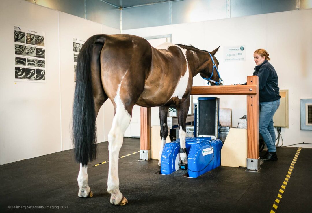
The horse remains standing during the scan, with only mild sedation required, thus eliminating any possible risks associated with general anesthesia. This makes sMRI a safe and repeatable option for diagnosing and monitoring the progression of equine injuries, without the harmful effects of radiation.
In fact, due to its excellent safety profile, sMRI is often preferred for repeat imaging. MRI rescans play a vital role in monitoring lesion progression, evaluating treatment responses and adjusting rehabilitation plans, including workload and therapy, based on both clinical and imaging findings.
Myth #2 – Standing Equine MRI Requires Surgery
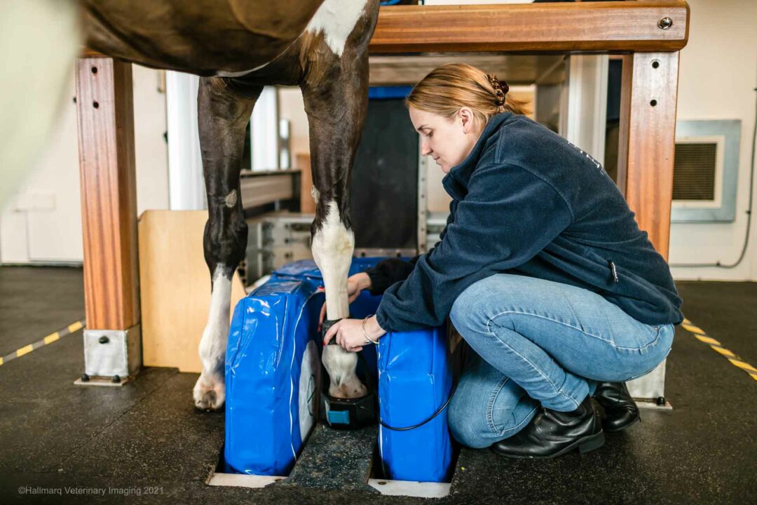
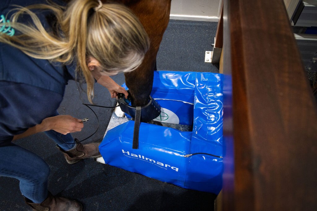
Another common myth is that an sMRI scan will require surgery. Not so! sMRI is a non-invasive imaging technique, meaning no incisions or invasive procedures are necessary to obtain images. Instead, the horse is gently sedated and walked into the MRI scanner, where the lame leg is carefully positioned between the magnet’s poles.
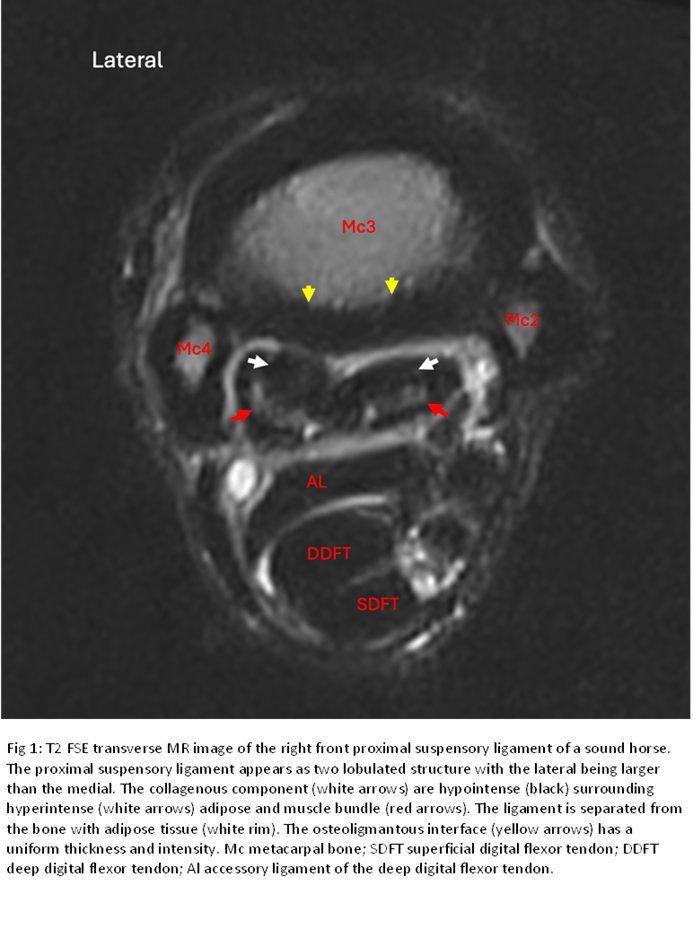
Proper positioning of the limb (Fig.2) is crucial to ensure that clinically diagnostic images are captured. This process provides vets with slice-by-slice grayscale images that allow for the detection of early structural and physiological changes in tissues, without requiring surgery or any other intrusive measures.
This makes sMRI especially useful for diagnosing soft-tissue and osseous abnormalities such as ligament and tendon injuries, osteoarthritis, bone edema and fractures particularly in areas like the foot, carpus and tarsus, where other imaging methods may miss subtle changes.
Once the MRI images are interpreted by a radiologist, then surgery of some form may well be the next step in some cases. However, the report would be discussed with both horse owner and referring veterinarian before a diagnosis and treatment plan are recommended and in the patient’s best interest.
Myth #3 – Standing Equine MRI is Only Used for Severe Cases and is too Expensive for Routine Use
While sMRI for horses is often associated with more severe lameness cases, it is not limited to diagnosing only life-threatening conditions such as penetrating injuries, fractures, septic joints etc. MRI is invaluable for early diagnosis in equine medicine, allowing veterinarians to detect injuries or developing conditions before they become clinically apparent. Unlike other imaging modalities, such as radiography, which may only reveal advanced lesions, sMRI can identify subtle changes in tissues, ligaments, tendons, and bones at their earliest stages.
These changes are often not detected by other imaging methods, or provide a more complete characterization of known pathological conditions. This ability to recognize pathologies before clinical signs appear can prevent conditions from worsening, enabling veterinarians to intervene sooner with more effective treatments. By diagnosing issues early, MRI offers horses the best chance for a successful recovery, often with less aggressive treatments and better overall outcomes.
It’s true that MRI scans can seem expensive, but this investment can ultimately save both time and money.
Early detection of conditions could mean less invasive treatments, potentially reduce the need for surgery, and enable faster recovery times, all of which can lower the overall cost of care in the long run. In fact, research shows (Koch et al., 2020) that horses diagnosed via MRI within the first 12 weeks of lameness onset are more likely to recover successfully, emphasising the importance of early intervention.
The detail that MRI can provide regarding extent, type, severity and, in some cases, chronicity of injury is useful from a diagnostic standpoint as well as for guiding therapeutic intervention. Understanding the extent of disease based on MRI examination can help more readily identify horses that are unlikely to be manageable with medical treatment and may benefit from surgery.
Myth #4 – Horses Need to be Anesthetized for Standing Equine MRI
Some horse owners worry that an MRI requires general anesthesia, which comes with inherent risks. However, this is not the case for many equine MRI scans. There are two main types of MRI systems used in equine medicine: low-field (0.1-0.5Tesla) and high-field (>1.5Tesla) MRI.
- Low-field MRI: can be performed either with the horse standing under mild sedation or under general anesthesia, depending on the specific clinical need.
- High-field MRI: (1.5 Tesla and above) requires the horse to be recumbent and imaged under general anesthesia, which presents increased risks, including complications during anesthesia that can be life-threatening for the horse.
While high-field MRI can offer improved image resolution, especially for detailed joint and cartilage assessments, low-field MRI still provides excellent diagnostic value, especially for soft tissue and bone pathology in many clinical scenarios (Bolen et al., 2010). The strength of the magnetic field, (measured in Tesla), plays a crucial role in image quality(Byrne et al., 2021), but it is not always necessary to opt for the highest field strength, especially when sMRI provides sufficient clarity and minimizes anesthesia risk.
Hallmarq’s Standing Equine MRI, a low-field MRI system, offers significant advantages by allowing the horse to remain standing with only mild sedation. This greatly reduces the risk of anesthesia-related complications, which occur in 1% of horses undergoing general anesthesia (Morgan et al., 2023). In addition, their award-winning motion correction software compensates for subtle movements of the sedated horse, ensuring that high-quality images can still be obtained even with slight motion. In fact, Hallmarq’s latest enhanced motion-correction technology, iNAV, enables faster scan times and improved diagnostic accuracy, making sMRI the safe, efficient, and cost-effective option for many lameness evaluations.
Myth #5 – Standing Equine MRI is too Stressful for Horses
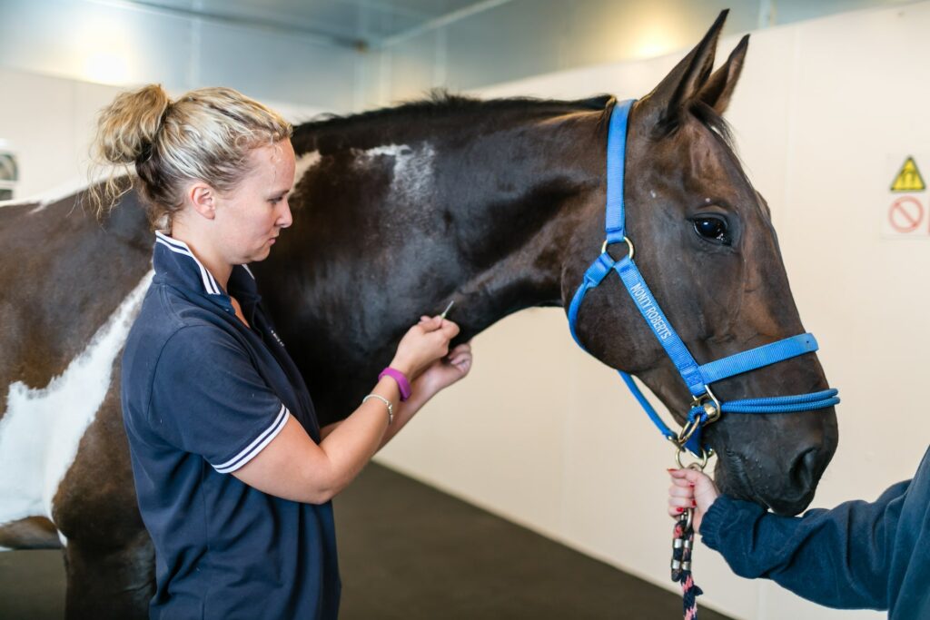
Horses are sensitive animals and it’s understandable that owners may worry about the stress associated with an sMRI scan. However, with the use of appropriate sedation and careful handling, horses are well prepared to remain calm throughout the procedure.
- A small amount of gentle sedation is administered to help the horse stay still, without the need for general anesthesia.
- An experienced veterinarian selects the appropriate sedation based on the horse’s health and the expected scan duration, ensuring both comfort and safety.
- Throughout the sMRI procedure, the horse’s vital signs are closely monitored to further minimize any risk or discomfort.
- Once sedated, the horse is walked into the sMRI scanner, and the lame leg is carefully positioned between the magnet’s poles.
- Proper limb positioning is essential for obtaining clinically diagnostic images.
- A radiofrequency (RF) coil is placed around the injury site to enhance image quality, and the operator ensures that both the patient and the sMRI system are correctly aligned.
With these careful measures in place, and with highly trained veterinarians and MRI technicians, the sMRI process remains safe, controlled, and as stress-free as possible for the horse.
Myth #6 – Standing Equine MRI Takes a Long Time
Another common belief is that sMRI scans are excessively time-consuming. While MRI might take longer than other imaging modalities such as radiographs and ultrasonography, the duration varies depending on:
- The area being scanned
- The number of regions examined
- The clinical questions raised
- The temperament of the horse
A single area, such as the foot, fetlock, or carpus, typically takes 45 to 60 minutes. However, veterinarians often request additional scans, such as the contralateral limb, which can extend the process.
Compared to other imaging techniques like nuclear scintigraphy, which takes at least two hours, sMRI is not significantly more time-consuming.
To ensure a stress-free experience, handlers take their time allowing the horse to adapt, with breaks between scans if needed. For example scanning both front feet typically takes 3 hours, and most horses can be seen on a day-patient basis. But if multiple areas are required, your vet may recommend that the horse stays overnight and be imaged over two days to avoid large amounts of sedation on one day.
For many horse owners, the benefits of obtaining high-quality, detailed diagnostic images far outweigh the time commitment involved in the procedure. For more information on the procedure, read our comprehensive guide:
How Hallmarq Supports Veterinary Practices With Equine MRI Technology
For over two decades, Hallmarq Veterinary Imaging has pioneered the field of equine lameness diagnosis. Their innovative standing equine MRI technology is considered the gold standard amongst equine veterinarians worldwide. Consistently at the forefront of veterinary imaging technology, Hallmarq is continually innovating to meet the needs of both vets and horse owners.
Hallmarq’s sMRI system is designed with both safety and efficiency in mind, offering high-quality images while minimizing stress for the horse. Their comprehensive training and ongoing support for veterinary practices is included in the company’s Q-Care Customer Programme enabling access to technology that helps deliver the highest standard of care to their patients.
Conclusion
Understanding the truth about sMRI is key to making informed decisions about your horse’s health. Contrary to popular myths surrounding Standing Equine MRI (sMRI), it is a safe, non-invasive, and effective diagnostic tool that can detect issues early, helping to prevent more serious problems down the line. It is neither dangerous, overly stressful, nor time-consuming, and while there are costs involved, the long-term benefits can far outweigh these initial expenses.
If your veterinarian recommends an sMRI procedure, you can be confident in the safety, effectiveness and value that this technology delivers. sMRI is a powerful tool in modern veterinary care, providing detailed insights that lead to better outcomes for your horse.
Always consult with your vet to determine whether an MRI is the best course of action for your horse’s specific condition, and don’t hesitate to ask questions to ensure you’re making the best choice for their long-term health.
References
- Bolen, G., Audigié, F., Spriet, M., Vandenberghe, F. and Busoni, V., 2010. Qualitative comparison of 0.27 T, 1.5 T, and 3T magnetic resonance images of the normal equine foot. Journal of Equine Veterinary Science, 30(1), pp.9-20.
- Byrne, C.A., 2021. Clinical magnetic resonance imaging of the equine foot: An investigation of factors influencing image quality and image interpretation (Doctoral dissertation, University of Glasgow).
- Koch DW, Barrett MF, Jackman BR, MacDonald D, Goodrich LR. (2020) Comparison of lameness outcomes in horses with acute or chronic digital lameness that underwent magnetic resonance imaging. N Z Vet J. 68(5):283-288. Morgan, Jessica M., Helen Aceto, Timothy Manzi, and Elizabeth J. Davidson. ‘Incidence and Risk Factors for Complications Associated with Equine General Anaesthesia for Elective magnetic resonance imaging’. Equine Veterinary Journal, 7 November 2023.