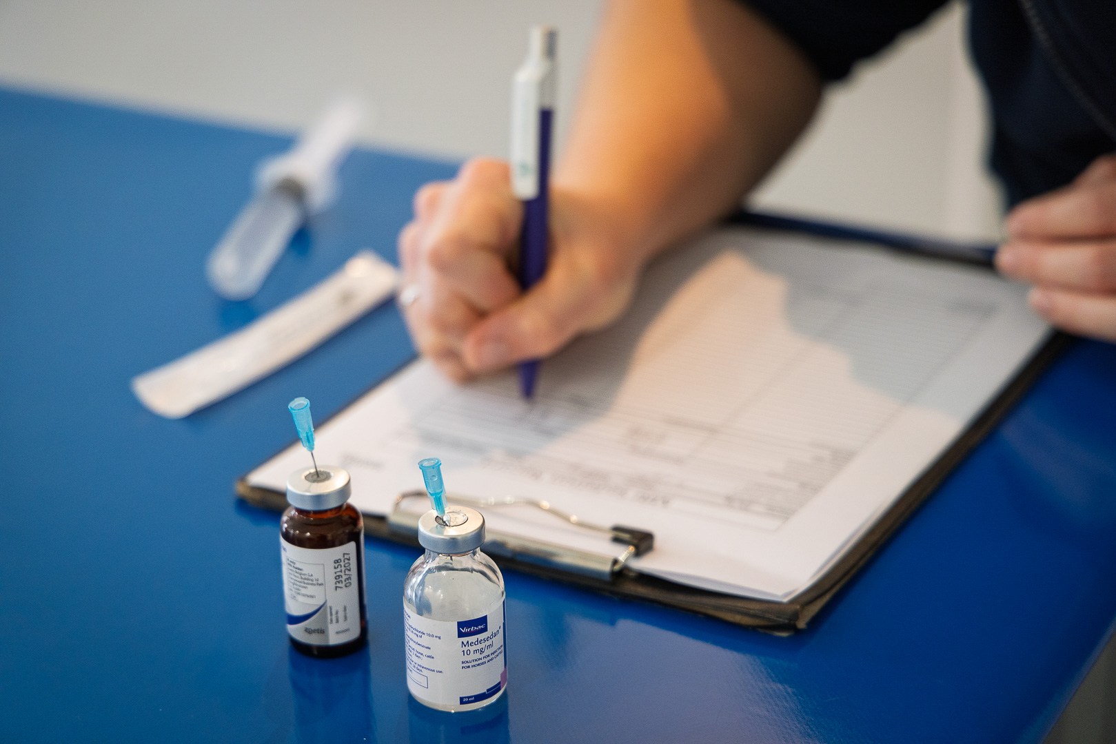




You’ve worked the case up, localized the lameness and decided that standing MRI is the sensible next step. For many referring veterinarians, that’s the point where a few practical questions surface and, sometimes, a quieter one underneath them all: what happens to my patient and my relationship with this client once I hand them over?
It’s a fair question and one worth answering plainly. Referral is not a handover of care. It’s a short, defined loan of your patient to a specialist facility for one purpose – a definitive diagnosis – after which the horse comes back to you with a treatment plan that you’ve helped shape.
This guide walks through exactly what happens from the moment you make the referral, to the moment the report lands, so you can refer with confidence and set your client’s expectations accurately.
Your Patient Stays Your Patient
The most important thing to know after you refer a horse for MRI is this: in every case, the horse is returned to your care. The referral center performs the scan, interprets the images and produces a report, but the ongoing treatment, monitoring and relationship remain with you.
There’s a long-standing worry among some referring veterinarians that sending a horse to a specialist means losing the client to that specialist. In practice, that fear is almost always unfounded and where it does happen, it runs against the ethical norms the whole referral system is built on. Referral centers depend on referring veterinarians to sustain their caseload; alienating the practices that send them work makes no sense commercially or professionally. The expectation across the profession is unambiguous – the patient goes back to the practitioner who knows them and the treatment plan is agreed between the referral center, you and the owner together.
Clients tend to value seeing their own veterinarian working alongside a specialist. That collaboration builds their confidence in the diagnosis and, in turn, improves compliance with whatever treatment follows. Far from weakening the relationship, a well-handled referral usually strengthens it.
Before the Scan: What the Referral Center Needs From You
Once you’re ready to refer a horse for MRI, most referral centers welcome a phone call before anything else. No two lameness cases are the same and a short conversation about the history and your findings helps the center plan the right protocol and the right regions to image.
It helps to have the following ready:
- The case history and your clinical findings – including the lameness pattern and how it has progressed.
- The results of your diagnostic analgesia. Localizing the source of pain before referral is essential. For a foot scan, the horse should have a positive response to a palmar digital or coffin joint block; an abaxial sesamoid block alone is not sufficient. Where a horse is positive to abaxial sesamoid but negative to more distal blocks, additional imaging of the pastern and fetlock is worth discussing.
- Previous imaging – any radiographs or ultrasound already acquired, which the specialist will read alongside the MRI rather than in place of it.
- Previous treatments and response – including any medication.
For non-urgent cases, the center will usually contact the owner directly to schedule an appointment. Owners should also be aware that horseshoes will need to be removed from the limb(s) being imaged. Arriving with clean, dry legs free from topical sprays or antiseptics helps produce the best possible MRI images.
On the Day: How the Horse is Scanned
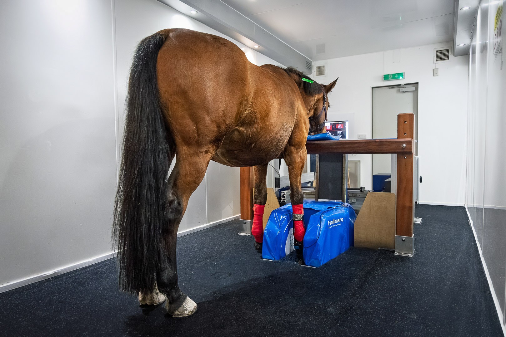
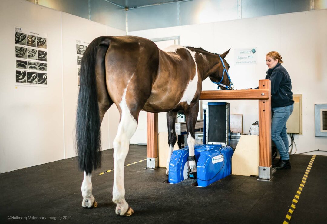
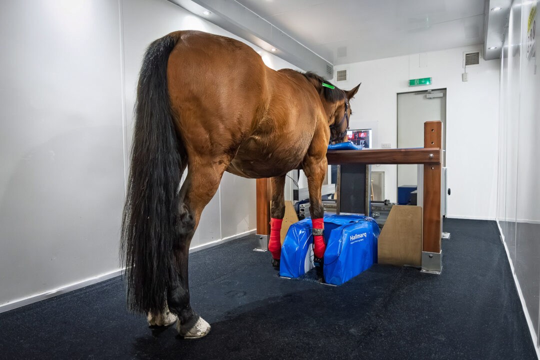
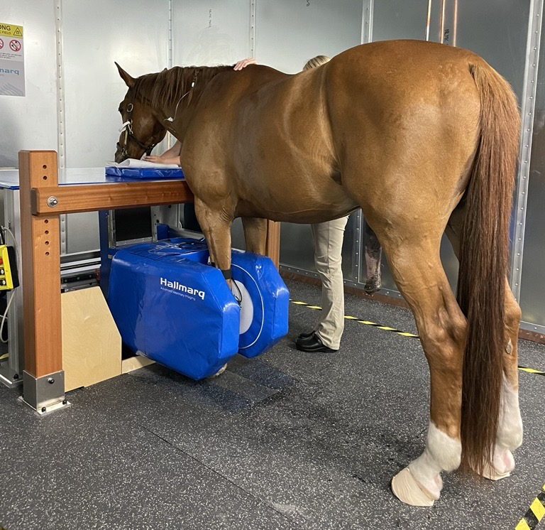
Reassurance for both you and your client here is straightforward, because the procedure is designed around the horse’s welfare. There is no general anesthesia. The horse stands, mildly sedated, with the limb positioned in the magnet inside a radiofrequency (RF) shielded room. No ionizing radiation is involved.
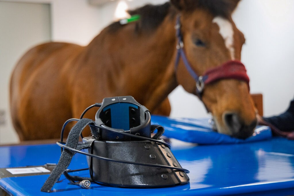

Careful positioning and a calm, well-prepared horse make all the difference to image quality. Even the most settled horse sways slightly when standing, so Hallmarq’s motion correction software – unique in the equine field – compensates for that movement and repeats any part of a sequence that would otherwise blur. A single region (foot or fetlock or PSL etc) can take anywhere between 20-45 mins depending on imaging protocol used and temperament of the horse. For patients where multiple regions of the limb need imaging, the time can add up. Occasionally the exam might be spread over two days to limit sedation. The majority of horses are seen as day patients and go home once they have recovered from sedation.
For a fuller walkthrough you can share with clients, the Guide to Standing Equine MRI covers preparation, the scan itself and aftercare in detail.
How the Report Comes Back to You
Once the scan is complete, a specialist interprets the images and produces a radiological report. A comprehensive report is usually available within a few days, with copies sent to both you and the owner.
The report does more than describe what was found. By arrangement, most centers will also provide:
- – The significance of the findings – which abnormalities are considered most relevant to the horse’s clinical signs and likely to be contributing to the lameness.
- – Prognosis and treatment recommendations – guidance on expected outcome and possible treatment options. If requested by the referring veterinarian, the report may also include a suggested treatment plan.
- – An explanation for the client in accessible language – to help you discuss the findings and their implications with the owner.
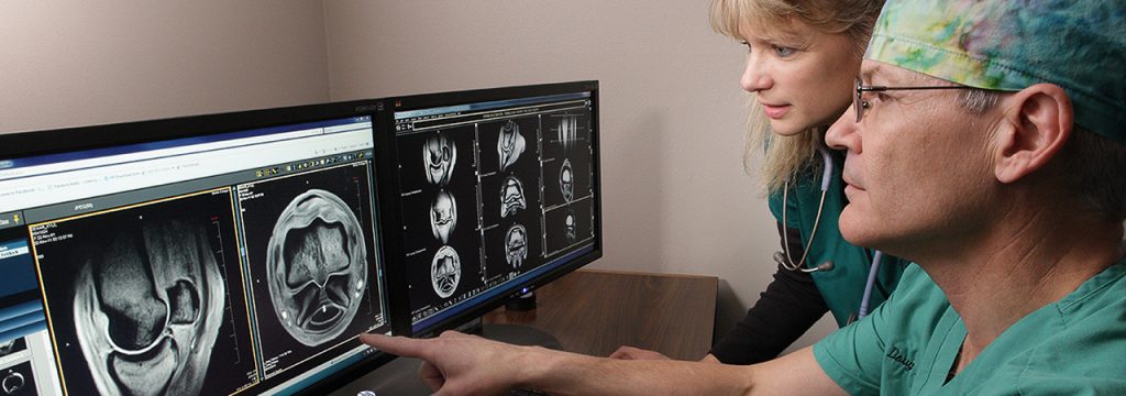
MRI’s value lies in what it reveals that other modalities can’t. It images bone and soft tissue in the same examination, distinguishes water from fat, and can identify early bone changes such as bone edema. It also helps assess the activity of lesions and their potential clinical significance, for example in tendons and ligaments, which may not be apparent from other imaging modalities.
Talking to the Owner
Because the report comes with a plain language explanation and the horse comes back to you, you’re well placed to lead the conversation that matters most: what the findings mean for this horse and what happens next.
The framing that tends to land is a collaborative one. You and a specialist have worked together to reach a definitive answer and that answer allows for a specific, targeted treatment rather than trial and error. Owners are increasingly familiar with MRI from human medicine and often arrive at the idea themselves; when they see their own veterinarian coordinating it, their confidence in the whole process grows.
Why the Referral is Worth Making
The case for early referral is a clinical one before it is a commercial one. Lameness that is treated without a clear diagnosis has a habit of returning – the horse comes back to the clinic again and again and each cycle of empirical treatment and rehabilitation carries its own cost in time, money and welfare. An early, definitive diagnosis breaks that loop.
In many horses, lameness is managed conservatively rather than surgically, but an accurate diagnosis is still essential to guide treatment and rehabilitation, establish a realistic prognosis, and understand what to expect for the horse’s future.
Horses that localize to the same region can have very different underlying pathology and imaging before committing to a procedure can confirm that surgery is the right call – or reveal pathology that changes the plan entirely and means conservative management is more appropriate. The alternative is discovering, twelve months and a full rehabilitation later, that the picture was never as simple as it looked. Our piece on whether to scan before surgery in proximal suspensory disease explores exactly this trade off. Seen in that light, the cost of a scan starts to look modest against the cost of getting the decision wrong.
Cases You Might Not Have Considered Scanning
It’s also worth revisiting what standing MRI can now see. Imaging is no longer confined to the foot – the distal limb can be assessed up to the carpus and hock, which brings a range of proximal limb cases into scope that might previously have been referred elsewhere or left undiagnosed. If it has been a while since you reviewed your own case selection, now is a good time to look again.
Making a Referral for Standing MRI
If your’e looking to refer a horse for MRI and have a case in mind, the simplest starting point is a conversation with your nearest facility. You can find a Hallmarq site here and the FAQs for equine vets answer the practical questions that come up most often. As ever, our team is happy to talk through anything you are unsure about.
Refer with confidence – the horse comes back to you and so does the client.
Explore our other articles: