A Substantial POD Lesion With Mild Uptake on Scintigraphy Images
History
A 3-year-old Thoroughbred colt had pulled up forelimb lame after his last race and subsequently developed a left hindlimb lameness. The cause of the forelimb lameness was identified using diagnostic analgesia and radiography, whilst the hindlimb lameness could not be localized.
Scintigraphy Findings
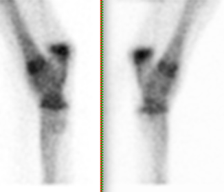
The colt underwent scintigraphic imaging of the lumbar spine, pelvis and hindlimbs to help identify the pathology present. No abnormalities were seen in the lumbar spine, pelvis, stifles and tibiae.
There was a linear increased radiopharmaceutical uptake bilaterally in the subchondral bone of the tarsometatarsal joints (figure 1).
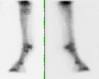
The plantarodistal aspect of the condyle of the left third metatarsal bones showed a mildly increased radiopharmaceutical uptake compared to the right (figure 2). This uptake was not visible in the plantar images and therefore laterality could not be determined.
CT Findings
The horse then underwent standing cone beam computed topography of the left hind fetlock joint prior to any further diagnostics. This displayed:
- A mild depression in the plantaro-distal articular surface of the lateral condyle of the third metatarsal bone.
- A separate ill-defined, bony fragment, seen at the level of the depression, measuring approximately 4.5mm in diameter and 1.6mm in thickness (figure 3).
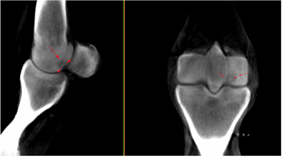
- A hypodense area involving the subchondral bone and trabecular bone deep to the abnormal articular surface (figure 3); this region was approximately 10mm in diameter and surrounded by a larger area with increased attenuation, consistent with sclerosis, affecting the entire plantar half of the lateral condyle.
- Mild subchondral bone thickening was also seen on the dorsal distal aspect of the sagittal ridge of the third metatarsal bone.
Conclusion
A conclusion was made that the horse showed severe plantar osteochondral disease (POD) with evidence of articular surface collapse affecting the lateral condyle of the third metatarsal bone which could explain the lameness observed. This lesion was suspected to correspond with the region of uptake detected scintigraphically and, because of the mild degree of uptake detected at this level, it was considered likely that the lesion was predominantly necrotic in nature, with only limited osteoblastic activity.
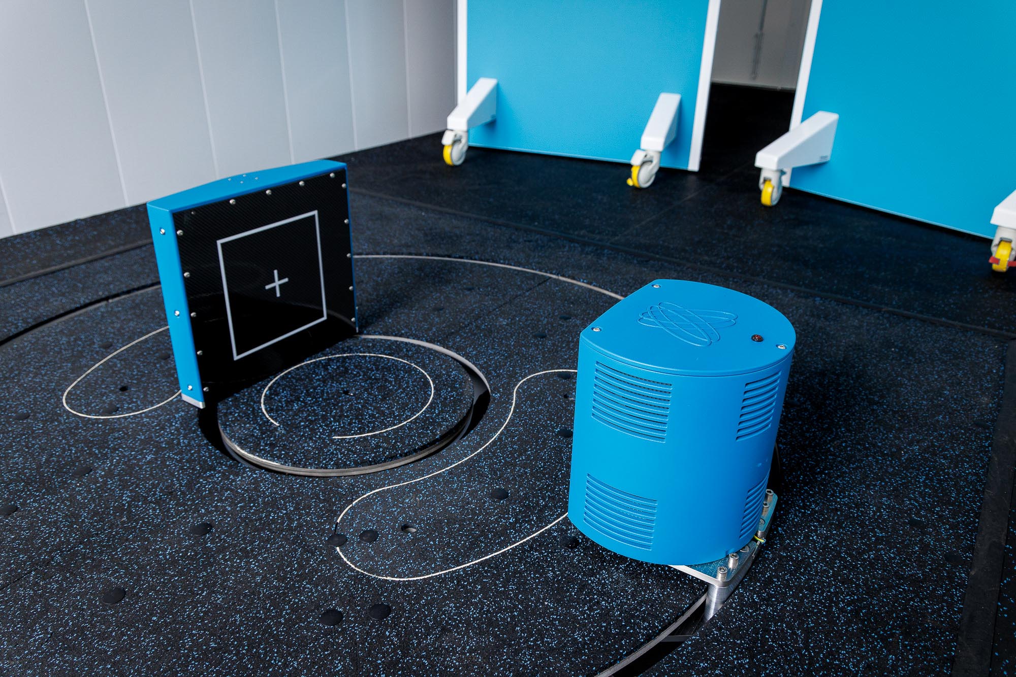
Access to Advanced Imaging for Your Patients
In this case study, CT scans were performed using Hallmarq’s Standing Equine Leg CT system. Images and interpretation provided in collaboration with VetCT.