

Cranial thoracic stenosis, a form of canine spinal stenosis, has often been studied in large breed and giant breed dogs such as German shepherds which are more commonly affected. However, the disease can appear in much smaller dogs, too, and specifically in screw-tail brachycephalic breeds.
Cranial thoracic stenosis is caused by a developmental malformation of the vertebral arch, specifically hypertrophy of both the lamina and the articular processes. It can occur unilaterally or bilaterally. This leads to a reduced dorsoventral or lateral diameter of the vertebral canal. Affected dogs typically show clinical signs of compressive myelopathy, characterised by non-painful progressive ataxia and paresis of the hind limbs.
We invited researcher and ECVS resident in small animal surgery Alessandro Conte to share his findings on cranial thoracic stenosis in screw-tail brachycephalic dogs (primarily French and English bulldogs).
Here’s what he’s learned regarding the disease pathogenesis, diagnosis and treatment.
Tracing the Disease Pathogenesis
Previous research has identified clinical, morphologic and morphometric features of clinical canine spinal stenosis in large and giant breeds. There are three potential presentations:
- Developmental: Present at birth and has an active cause that is present during the growth period.
- Acquired: Comes about as a result of ligamentous hypertrophy, degenerative articular changes or, synovial cysts.
- Congenital: Present at birth, but with no active underlying cause (most likely genetic).
The developmental etiology, previous authors have shown, is the most likely explanation for cranial thoracic spinal stenosis because the clinical signs were present early, even before skeletal maturity.
You can trace the disease pathogenesis through 3D imaging. In healthy dogs, the angles formed by articular processes are shallow. But in dogs with canine spinal stenosis, the articular processes are much more angled. These abnormalities of articular geometry put additional stressors on the joint and likely play a large role in the development of spinal stenosis.
Imaging and Diagnosing Canine Spinal Stenosis
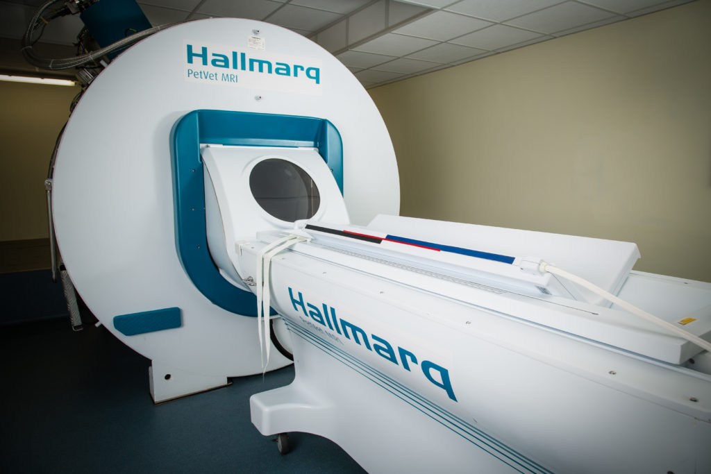
MRI is the gold standard for imaging spinal diseases. To get the best images, your MRI study should consist of at least a sagittal, a transverse and a dorsal T2-weighted sequence as well as, ideally, a T1-weighted sequence. These sequences should be taken from the cranial thoracic to the mid-lumbar region.
There are advantages and disadvantages of using MRI to diagnose canine spinal stenosis:
- Advantages: MRI provides superior soft tissue resolution and allows you to acquire much more information on spinal cord integrity and pathology.
- Disadvantages: It can be very costly for both clients and practitioners. For that reason, MRI may be difficult to access in your local area. Additionally, older dogs with pacemakers or metallic implants may not be qualified for an MRI study.
Computed tomography (CT) is an alternative imaging tool for diagnosing canine spinal stenosis. Like MRI, CT scans should also capture sagittal, transverse and dorsal sequences, with a contrast medium for the best results. These images allow for a 3D construction of the study so you can isolate only the affected vertebrae.
CT also presents unique advantages and disadvantages.
- Advantages: CT scans provide superior bone tissue resolution. CT tends to be non-invasive (whether plain or contrast-enhanced), and it’s often more readily available than MRI.
- Disadvantages: Intrathecal contrast is very much required for the identification of some pathologies. CT scans provide little information on the integrity or pathology of the spinal cord.
Diagnosis for a Clear Picture
While MRI is ideal for diagnosing the disease, CT can also produce diagnostic-quality images. And if you have access to both options, you can get a clear picture of your patient’s condition.
Once your scans are complete, find where the spinal cord appears to be most compressed. By calculating the conceptual surface area, you can get a sense of where the compressive myelopathy is most severe.
Options for Treating the Disease
There’s little information in the literature on treating screw-tail brachycephalic dogs for canine spinal stenosis. However, one case report reveals that a patient underwent a dorsal laminectomy to surgically relieve pressure on the vertebral canal, doing so with a successful outcome. Similar procedures have produced good outcomes in other (non-brachycephalic) breeds, too.
Medical management can also produce a fair outcome. But while their prognosis is good, these patients will continue to have neurological deficits.
Patients included in Conte’s study tended to respond well to either surgical treatment or medical management, but the limited number of dogs in the study and the absence of follow-up information prevents him as yet from being able to draw conclusions regarding the long term outcome of canine spinal stenosis in screw-tail brachycephalic dogs.
There’s still a lot more to discover about canine spinal stenosis. Watch the complete webinar with Conte for more information.